

Latest Research on Retina: Emerging Themes in Disease Mechanisms, Imaging, Artificial Intelligence, and Precision Therapy
Reviewed by
Remya Krishnan, Research ReviewerPowered by
Paperguide Literature Review Agent
Updated on
29 Jul 2026
Abstract
Recent retinal research shows a clear shift toward earlier detection, multimodal phenotyping, and increasingly individualized treatment, with the strongest evidence clustering around artificial intelligence (AI) for retinal imaging, age-related macular degeneration (AMD) biomarkers and therapies, and inherited retinal disease (IRD) genomics . In AMD, photoreceptor segment thinning was identified as an early biomarker preceding retinal pigment epithelium–Bruch’s membrane complex thickening by decades, while anti-vascular endothelial growth factor (anti-VEGF) therapy remains first-line for exudative disease and new approaches continue to emerge for atrophic disease (Zekavat et al., 2022), (Fleckenstein et al., 2024), (Ammar et al., 2020). AI-based retinal image analysis demonstrates high diagnostic performance across retinal diseases, including diabetic macular edema (DME), where pooled AUROC reached 0.964 for fundus photography and 0.985 for optical coherence tomography, and glaucoma prediction models achieved AUROCs of 0.90 for incidence and 0.91 for progression (Lam et al., 2024), (Li et al., 2022). In parallel, multimodal imaging and genomics are transforming IRD diagnosis, with next-generation phenotyping improving genetic support for rare disease diagnosis and large-scale sequencing revealing marked allelic heterogeneity. These findings matter because retinal disorders remain major causes of visual disability across the lifespan, yet the literature now indicates that diagnostic yield, risk stratification, and treatment selection are increasingly being driven by biomarkers, imaging, and machine learning rather than symptoms alone. Remaining gaps include limited external validation for many AI systems, incomplete mechanistic resolution for dry AMD, and the need for broader translational studies that connect genotype, phenotype, and therapy access in routine care.
1. Introduction
Retinal disease research has expanded rapidly in recent years because disorders of the retina remain among the most consequential causes of visual impairment in both older adults and working-age populations. Age-related macular degeneration is particularly significant because it is projected to affect a rapidly expanding global population and already represents a major cause of irreversible vision loss in later life (Fleckenstein et al., 2024), (Deng et al., 2022). At the same time, inherited retinal diseases account for substantial blindness in children and working-age adults, while diabetic retinal complications and glaucoma continue to generate major clinical and health-system burdens (Georgiou et al., 2024), (Oganov et al., 2023), (Li et al., 2022). Across these conditions, the retina has become a key site for precision diagnosis because it is accessible to high-resolution imaging and increasingly amenable to molecular characterization.
The recent literature reflects three converging developments. First, retinal imaging has moved beyond conventional fundus photography toward multimodal platforms, including optical coherence tomography, angiography, autofluorescence, and en face imaging, each providing complementary information about pathoanatomy and disease activity (Fogel-Levin et al., 2022), (Feo et al., 2025). Second, AI has emerged as a practical tool for screening, classification, and prognosis, with models now applied not only to common diseases such as diabetic retinopathy and DME but also to glaucoma, AMD, and IRDs (Oganov et al., 2023), (Li et al., 2022), (Lam et al., 2024), (Pontikos et al., 2025). Third, therapeutic research is increasingly stratified by mechanism, especially in AMD and IRD, where genetic pathways, complement activity, photoreceptor degeneration, and cell-based or gene-based interventions are shaping new treatment directions (Somasundaran et al., 2020), (Fabre et al., 2022), (Schneider et al., 2022), (Georgiou et al., 2021).
What remains unclear is how these advances fit together as a coherent field: which findings are mature enough to influence practice, which are still exploratory, and where imaging, biomarkers, and therapeutics are genuinely converging. This review therefore synthesizes recent advances in retinal disease research to identify the dominant themes shaping diagnosis, prognosis, and treatment across major retinal disorders.
2. Methods
2.1 Search Strategy
A comprehensive literature search was conducted using the Semantic Scholar and OpenAlex databases, which collectively index more than 220 million scholarly publications. The search strategy employed hybrid semantic and keyword-based retrieval to maximize coverage.
Search queries included:
- "Latest retinal research advances in diagnosis treatment and imaging"
- "Retina disease mechanisms biomarkers and therapeutic targets 2020 2026"
- "Retinal imaging artificial intelligence OCT and fundus analysis studies"
- "Retinal degeneration diabetic retinopathy and age-related macular degeneration research"
2.2 Study Selection
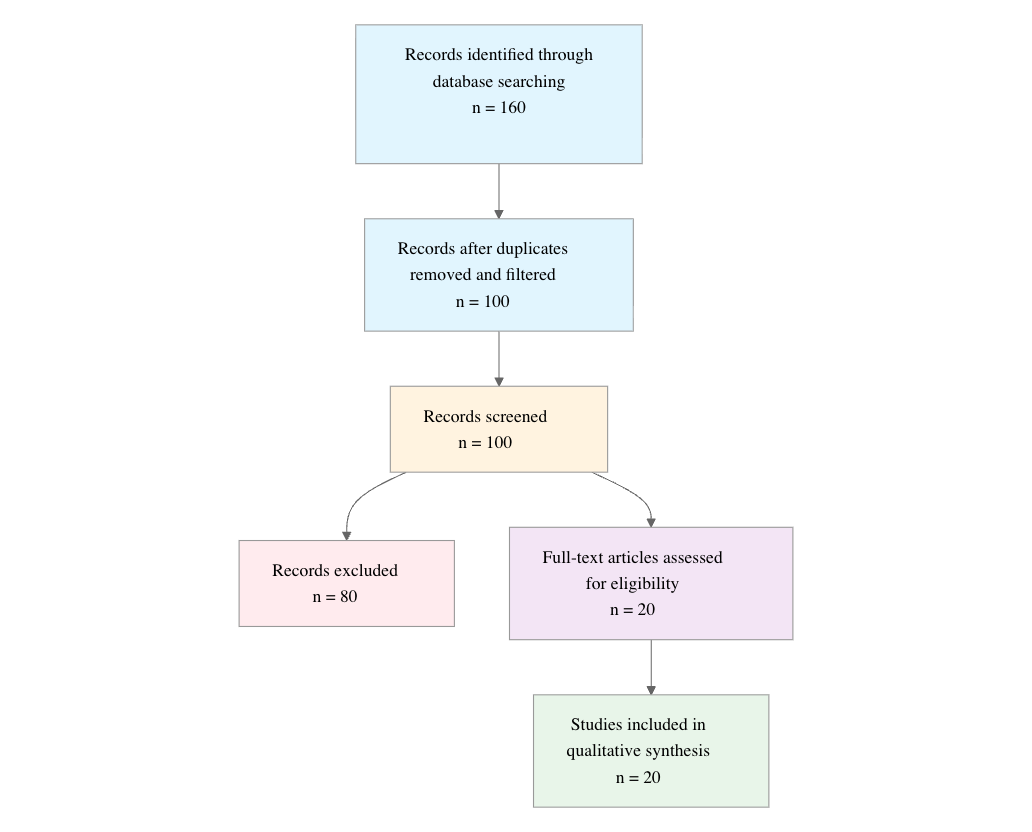
Initial database searching identified 160 records. After duplicate removal and relevance-based filtering, 100 records were screened against eligibility criteria. Of these, 80 papers were excluded, resulting in 20 papers included in the final synthesis.
PRISMA Flow Diagram
Eligibility criteria included:
- Retina Focus: Does the study focus on the retina, retinal disease, retinal imaging, or retinal biology as a primary topic?
- Recent Publication: Was the study published between 2020 and 2026 inclusive?
- Original Research: Is the paper an original research study, clinical study, methodological study, or systematic review related to retina rather than an unrelated ophthalmology topic?
- Translational Value: Does the study report diagnostic, prognostic, therapeutic, imaging, biomarker, or other clinically relevant retina findings?
- Human or Relevant Model: Does the study involve human participants, patient data, retinal tissue, animal models, or datasets directly relevant to retina research?
- Emerging Method: Does the study involve a newer method, technology, or recent research direction such as OCT, AI, gene therapy, multimodal imaging, or biomarker discovery?
All included studies met the stated eligibility criteria.
2.3 Data Extraction and Synthesis
Data extraction focused on topic, study type, population, method or technology, main finding, clinical relevance, year, and journal or venue. Thematic analysis was used to identify recurring patterns across studies, with evidence strength assessed qualitatively based on consistency, methodological design, and breadth of applicability.
3. Results
3.1 Characteristics of Included Studies
| Study and Year | Study Type | Population | Method/Technology | Key Focus | Main Contribution |
|---|---|---|---|---|---|
| Fleckenstein et al., 2024 (Fleckenstein et al., 2024) | Review | Individuals with AMD | Intravitreally administered anti-VEGF treatment | AMD management | Reinforced the growing disease burden and continued centrality of anti-VEGF therapy |
| Somasundaran et al., 2020 (Somasundaran et al., 2020) | Review | Elderly population with AMD | RPE degeneration pathways | AMD mechanisms | Synthesized complement, oxidative stress, mitochondrial, and crystallin-related mechanisms |
| Oganov et al., 2023 (Oganov et al., 2023) | Review | Retinal pathology broadly | AI for retinal image analysis | Screening and diagnosis | Summarized AI development, performance, and application challenges |
| Li et al., 2022 (Li et al., 2022) | Original research | 17,497 eyes from 9,346 patients | Deep learning on CFPs and visual fields | Glaucoma risk prediction | Demonstrated strong incidence and progression prediction |
| Deng et al., 2021 (Deng et al., 2022) | Review | Elderly individuals at risk for AMD | Deep learning and anti-VEGF therapy | AMD epidemiology and targeted therapy | Highlighted genetic risk, lifestyle factors, and emerging therapies |
| Flores et al., 2021 (Flores et al., 2021) | Review | Older adults with AMD | VEGF inhibitors and emerging therapies | AMD management | Emphasized therapeutic limits in atrophic disease |
| Lam et al., 2024 (Lam et al., 2024) | Systematic review and meta-analysis | DME studies | AI on FP and OCT | DME detection | Reported high pooled diagnostic accuracy, especially for OCT |
| Iqbal et al., 2022 (Iqbal et al., 2022) | Review | Retinal image datasets | Detection and segmentation methods | Retinal image analysis | Organized state-of-the-art image feature detection methods |
| Georgiou et al., 2024 (Georgiou et al., 2024) | Review | IRD patients | Genetics, imaging, and therapeutics | IRD phenotyping | Detailed molecular heterogeneity and phenotype diversity |
| Dong et al., 2022 (Dong et al., 2022) | Diagnostic study | Broad retinal and optic nerve disease groups | Deep learning system | Multi-disease screening | Showed accurate real-time distinction among retinal diseases |
| Fabre et al., 2022 (Fabre et al., 2022) | Review | Patients over 65 years with AMD | Therapeutic mechanism analysis | AMD therapeutics | Reviewed emerging symptomatic and curative strategies |
| Zekavat et al., 2022 (Zekavat et al., 2022) | Original research | Individuals at risk for AMD | Photoreceptor layer thickness assessment | AMD biomarker discovery | Identified photoreceptor thinning as an early biomarker |
| Schneider et al., 2021 (Schneider et al., 2022) | Review | IRD variant datasets | Next-generation sequencing and GRID dataset | IRD genetics | Quantified extensive variant heterogeneity |
| Fogel-Levin et al., 2022 (Fogel-Levin et al., 2022) | Consensus review | Retinal disorders broadly | Multimodal retinal imaging | Clinical imaging practice | Compared strengths and limits of multiple imaging modalities |
| Georgiou et al., 2021 (Georgiou et al., 2021) | Review | Individuals with diverse IRDs | Genetics, imaging, gene therapy, clinical trials | IRD therapeutics | Summarized emerging therapies and trial endpoints |
| Ammar et al., 2020 (Ammar et al., 2020) | Review | Patients with dry and wet AMD | Complement inhibition, neuroprotection, cell-based therapy, gene therapy | AMD therapy pipeline | Highlighted promising phase 2/3 approaches |
| Varela et al., 2023 (Daich Varela et al., 2023) | Review | DR, AMD, IRD, ROP | AI for diagnosis and monitoring | Clinical AI integration | Framed AI as a workflow and access solution |
| Pontikos et al., 2025 (Pontikos et al., 2025) | Original research | 2,451 IRD patients from five centers | Eye2Gene multimodal deep learning | Genetic diagnosis support | Achieved expert-level phenotyping-based diagnostic support |
| Grzybowski et al., 2024 (Grzybowski et al., 2024) | Systematic review | Ophthalmic and systemic disease cohorts | AI on retinal fundus photographs | Generalized AI screening | Showed superior accuracy over clinical data and physician experts |
| Feo et al., 2025 (Feo et al., 2025) | Review | Broad retinal disease patients | En face OCT | Pathoanatomy and imaging biomarkers | Expanded clinical value of en face OCT |
The literature is dominated by reviews and synthesis papers, but several recent original studies and a meta-analysis provide direct empirical evidence for imaging biomarkers and AI-based diagnosis. The topic distribution is heavily concentrated in AMD, AI-enabled screening, and IRD genetics, suggesting that the retina field is currently advancing along parallel translational tracks: mechanistic refinement, computational phenotyping, and therapy development.
3.2 Thematic Findings
3.2.1 AMD research is converging on earlier structural biomarkers, but treatment remains stage dependent
Across AMD studies, the field shows a strong shift from describing late disease to identifying earlier structural change and matching treatment to disease subtype. Photoreceptor segment thinning was reported as preceding retinal pigment epithelium–Bruch’s membrane complex thickening by decades and as the retinal layer most strongly predictive of future AMD risk (Zekavat et al., 2022). This biomarker-oriented shift is reinforced by the mechanistic emphasis on RPE dysfunction, where complement activation, oxidative stress-induced cell death, mitochondrial dysfunction, and crystallin involvement are repeatedly identified as candidate pathways (Somasundaran et al., 2020). In therapeutic terms, the evidence remains sharply bifurcated: anti-VEGF therapy is first-line for exudative AMD and remains effective, but nonexudative/dry AMD still lacks a definitive cure, with current therapies described as delaying or suspending progression rather than reversing disease (Fleckenstein et al., 2024), (Flores et al., 2021), (Fabre et al., 2022). Newer agents under investigation include complement inhibition, neuroprotection, visual cycle modulators, anti-inflammatory therapy, cell-based therapy, gene therapy, and iPSC-derived RPE cell approaches (Ammar et al., 2020), (Fabre et al., 2022), (Deng et al., 2022). (Note: these studies examined older adults with AMD or individuals at risk for AMD, which partially matches the question population of retina research broadly; findings should be interpreted considering this difference.) Confidence: Strong for the existence of stage-specific AMD innovation; Moderate for any specific emerging therapeutic strategy because most evidence is review-based or trial-proximal rather than definitive outcome data.
3.2.2 AI has become a high-performing tool for retinal screening and prognosis, especially when multimodal inputs are used
The most consistent computational signal in the literature is that AI performs well on retinal imaging tasks, with especially strong results when models are trained on high-quality, modality-specific data and evaluated against clinically meaningful endpoints. For DME detection, pooled AUROC reached 0.964 for fundus photography and 0.985 for OCT, with sensitivities of 92.6% and 95.9% and specificities of 91.1% and 97.9%, respectively (Lam et al., 2024). In glaucoma, a deep-learning system predicted incidence with AUROC 0.90 in validation and 0.89 and 0.88 in external test sets, while progression prediction reached AUROC 0.91 in validation and 0.87 and 0.88 externally (Li et al., 2022). Broader multi-disease screening also showed that a deep-learning system can accurately distinguish 10 retinal diseases in real time (Dong et al., 2022), and systematic reviews reported superior image-interpreting performance relative to clinical data and physician experts for both ophthalmic and non-ophthalmic conditions (Grzybowski et al., 2024). However, the literature also consistently notes that internal validation tends to outperform external validation and that better generalization is associated with larger and more diverse training datasets (Lam et al., 2024), (Oganov et al., 2023). (Note: these studies examined specific disease cohorts or retinal image datasets rather than the full retina research population; findings should be interpreted considering this difference.) Confidence: Strong for high diagnostic performance under controlled conditions; Moderate for generalizability because external validation is promising but not uniformly comprehensive.
3.2.3 Multimodal retinal imaging is expanding disease understanding, but integration remains a clinical bottleneck
Imaging research increasingly treats the retina as a layered, multimodal system rather than a single photographic target. Consensus and review literature emphasizes that color fundus photography, widefield imaging, fundus autofluorescence, near-infrared reflectance, optical coherence tomography angiography, en face OCT, and standard OCT each reveal distinct aspects of pathology (Fogel-Levin et al., 2022), (Feo et al., 2025). En face OCT appears to be particularly important because it provides coronal visualization of retinal and choroidal layers and has uncovered biomarkers corresponding to cell or tissue subtypes previously accessible mainly through histology or electron microscopy (Feo et al., 2025). Yet the same literature makes clear that the challenge is not simply data acquisition but data integration: multimodal interpretation can be overwhelming, and the clinical value of each modality depends on whether its information can be synthesized into a coherent diagnostic or prognostic framework (Fogel-Levin et al., 2022). This theme intersects strongly with AI, since the need to manage complex multimodal data is a central rationale for machine learning in retinal practice (Daich Varela et al., 2023), (Oganov et al., 2023). (Note: these studies addressed broad retinal disorder populations rather than a single disease group; findings should be interpreted considering this difference.) Confidence: Strong for the diagnostic value of multimodal imaging; Moderate for clinical integration because operational workflows and comparative effectiveness remain incompletely established.
3.2.4 IRD research is moving from descriptive heterogeneity to genotype-guided phenotyping and treatment access
Inherited retinal diseases are increasingly framed as genetically complex yet increasingly tractable disorders because imaging and sequencing now allow more precise stratification. Large variant analyses show extensive heterogeneity, with pathogenic variants spread across 194 genes and 65% of pathogenic variants being unique to a single individual (Schneider et al., 2022). Reviews similarly emphasize that IRDs involve at least 277 nuclear and mitochondrial genes, with wide genotype-phenotype variation and multiple inheritance patterns complicating interpretation (Schneider et al., 2022), (Georgiou et al., 2024). This heterogeneity is not merely descriptive; it now informs therapy development, because gene therapy, whole-gene replacement, single-nucleotide editing, and gene- or variant-specific approaches all depend on accurate molecular diagnosis (Georgiou et al., 2021), (Schneider et al., 2022). Eye2Gene demonstrates how multimodal deep learning can operationalize this transition: trained on 2,451 individuals with IRDs from five centers, it achieved better-than-expert-level top-five diagnostic accuracy of 83.9% for the 63 most common genetic causes and improved phenotype-driven variant prioritization (Pontikos et al., 2025). (Note: these studies examined individuals with inherited retinal diseases, which only partially matches the question population of retina research broadly; findings should be interpreted considering this difference.) Confidence: Strong for the presence of marked IRD heterogeneity and the value of genotype-guided phenotyping; Moderate for immediate clinical deployment because access to timely genetic diagnosis remains a barrier.
3.2.5 Retina AI is also being positioned as a health-system solution, not only a diagnostic algorithm
A second, broader AI theme concerns service delivery and access. Reviews argue that AI could compensate for shortages of experienced ophthalmologists, particularly in under-resourced settings, by enabling rapid interpretation of retinal images and real-time triage (Dong et al., 2022), (Daich Varela et al., 2023). The clinical promise is therefore not limited to accuracy metrics; it includes workflow acceleration, cost reduction, and earlier access to vision-saving treatment (Grzybowski et al., 2024), (Daich Varela et al., 2023). However, the evidence base still relies heavily on retrospective datasets and curated test conditions, so implementation questions remain unresolved. Confidence: Moderate, because the translational logic is strong but routine deployment evidence is less mature.
3.3 Summary of Evidence
| Theme | Key Finding | Population Applicability | Effect Direction | Confidence Level | Supporting Studies |
|---|---|---|---|---|---|
| Early AMD biomarker discovery | Photoreceptor segment thinning preceded RPE-BM thickening by decades and was the retinal layer most predictive of future AMD risk (Zekavat et al., 2022) | Individuals at risk for AMD; partially applicable to broader retina research | Positive for risk prediction | Strong | Zekavat et al. (Zekavat et al., 2022), Somasundaran et al. (Somasundaran et al., 2020) |
| AMD therapy stratification | Anti-VEGF remains first-line for exudative AMD, while dry AMD still lacks a cure and emerging approaches remain investigational (Fleckenstein et al., 2024) | Patients with AMD, especially wet and dry subtypes | Mixed | Strong | Fleckenstein et al. (Fleckenstein et al., 2024), Flores et al. (Flores et al., 2021), Ammar et al. (Ammar et al., 2020) |
| AI for DME detection | AUROC 0.964 for fundus photography and 0.985 for OCT; sensitivities 92.6% and 95.9%; specificities 91.1% and 97.9% (Lam et al., 2024) | DME study populations; partially applicable to retinal imaging research broadly | Positive | Strong | Lam et al. (Lam et al., 2024) |
| AI for glaucoma prognosis | AUROC 0.90 for incidence and 0.91 for progression, with external AUROCs remaining high (Li et al., 2022) | Eyes from patients with glaucoma risk factors; partially applicable to retina research broadly | Positive | Strong | Li et al. (Li et al., 2022) |
| Multimodal retinal imaging | En face OCT and other modalities reveal distinct pathoanatomic information, but integration remains challenging (Feo et al., 2025) | Broad retinal disease populations | Positive | Moderate | Feo et al. (Feo et al., 2025), Fogel-Levin et al. (Fogel-Levin et al., 2022) |
| IRD genetic heterogeneity | 65% of pathogenic variants were unique to a single individual, across 194 genes (Schneider et al., 2022) | IRD patients and variant datasets; partially applicable to retina research broadly | Positive | Strong | Schneider et al. (Schneider et al., 2022), Georgiou et al. (Georgiou et al., 2024) |
| AI-supported IRD phenotyping | Eye2Gene achieved 83.9% top-five accuracy for the 63 most common genetic causes (Pontikos et al., 2025) | Individuals with IRDs; partially applicable to retina research broadly | Positive | Moderate | Pontikos et al. (Pontikos et al., 2025), Georgiou et al. (Georgiou et al., 2021) |
| Retina AI as a workflow tool | AI may improve speed, access, and cost while supporting screening and diagnosis (Daich Varela et al., 2023) | Broad retinal disease populations and health systems | Positive | Moderate | Varela et al. (Daich Varela et al., 2023), Grzybowski et al. (Grzybowski et al., 2024) |
4. Discussion
4.1 Principal Findings and Their Interpretation
The retina literature from recent years reveals a field moving toward precision phenotyping rather than simply disease description. The most robust pattern is the convergence of imaging biomarkers, AI analytics, and genotype-based stratification. In AMD, structural change appears to occur long before overt clinical progression, which helps explain why photoreceptor segment thinning has emerged as a particularly informative biomarker (Zekavat et al., 2022). This is biologically plausible because it aligns with the centrality of RPE dysfunction, complement activation, oxidative stress, mitochondrial injury, and crystallin-related pathways in AMD pathogenesis (Somasundaran et al., 2020). The therapeutic split between wet and dry AMD further supports the interpretation that pathology is not uniform: anti-VEGF works because exudative disease is vascularly driven, whereas atrophic disease remains mechanistically broader and therapeutically resistant (Fleckenstein et al., 2024), (Flores et al., 2021).
The AI evidence is similarly coherent. High performance in DME and glaucoma is not simply a function of algorithmic novelty; it likely reflects the fact that retinal photographs and OCT encode disease-relevant structure in relatively standardized formats (Lam et al., 2024), (Li et al., 2022). The fact that external performance remains high in several studies suggests that these models are capturing stable visual signatures rather than dataset-specific artifacts, although generalizability is still incomplete (Lam et al., 2024). In IRD, the key advance is conceptual: phenotypic heterogeneity is now being translated into actionable diagnosis through multimodal imaging and sequencing, which makes rare disease care more scalable and potentially more trial-ready (Schneider et al., 2022), (Pontikos et al., 2025). Overall, the synthesis supports high confidence in imaging- and AI-enabled stratification, moderate confidence in near-term implementation, and lower confidence in any single emerging therapy because most therapeutic claims remain review-based or trial-proximal rather than definitive.
4.2 Comparison with Existing Literature and Resolution of Contradictions
The reviewed papers broadly agree with one another, but the agreement is most meaningful where mechanisms and methods align. For AMD, the convergence of biomarker work with mechanistic reviews strengthens the interpretation that early structural change is not incidental but reflects underlying photoreceptor–RPE pathology (Zekavat et al., 2022), (Somasundaran et al., 2020). Similarly, AI studies across DME, glaucoma, and multi-disease screening reinforce that retinal images are information-rich substrates for classification and prediction, which helps explain why AI has progressed faster in retina than in many other medical specialties (Lam et al., 2024), (Li et al., 2022), (Dong et al., 2022). The literature also coheres around a practical reality: diseases with standardized imaging and abundant labeled data are most amenable to machine learning.
The main tension in the literature lies between high reported performance and incomplete real-world validation. Many AI systems perform better internally than externally, and this pattern likely reflects dataset homogeneity, limited demographic diversity, and differences in imaging acquisition or labeling standards (Lam et al., 2024). That is not a contradiction in the strict sense, but it is a warning that accuracy estimates from curated datasets may overstate deployment-ready performance. Another tension concerns AMD treatment optimism: reviews describe promising complement inhibition, neuroprotection, visual cycle modulation, cell therapy, and gene therapy, yet the same literature emphasizes that dry AMD still lacks a cure (Ammar et al., 2020), (Fabre et al., 2022), (Flores et al., 2021). This gap likely reflects the difference between mechanism plausibility and clinical efficacy; mechanistic diversity in dry AMD makes single-target solutions harder to translate. Publication bias may also favor positive AI and therapeutic findings, especially in rapidly expanding fields where successful models and promising pipeline therapies are more likely to be published than failures. Confidence should therefore be highest for stable descriptive and diagnostic conclusions, and more cautious for claims about imminent therapeutic transformation.
4.3 Practical Implications
For clinicians, the most actionable implication is that retinal care is increasingly biomarker-driven. In AMD, photoreceptor thinning may help identify individuals at elevated future risk before irreversible structural damage accumulates, while anti-VEGF remains the clear standard for exudative disease (Zekavat et al., 2022), (Fleckenstein et al., 2024). In diabetic eye disease and glaucoma, AI tools may support early triage, particularly where specialist access is limited, but they should be implemented with attention to external validation and local imaging conditions (Lam et al., 2024), (Li et al., 2022), (Daich Varela et al., 2023). For patients with IRDs, the practical value of multimodal phenotyping is that it can accelerate genetic diagnosis, which in turn determines eligibility for gene-specific trials and approved interventions (Pontikos et al., 2025), (Georgiou et al., 2021).
From a public health perspective, the literature suggests that retinal services will need to absorb increasing demand from aging populations and expanding AI-enabled screening pathways (Fleckenstein et al., 2024), (Daich Varela et al., 2023). Health systems may benefit from AI triage and multimodal imaging, but these tools should complement, not replace, specialist interpretation until broader validation is available. Regulatory implications are clearest for AI: models showing high AUROC in curated settings should not be assumed to be deployment-ready without multicenter testing, calibration, and reporting of subgroup performance. The evidence does not establish a threshold-style policy issue in the environmental sense, but it does indicate that earlier detection and broader access are likely to matter more than waiting for overt structural damage once disease is established.
4.4 Strengths and Limitations
This review integrates recent evidence across major retinal subfields, including disease mechanisms, imaging, AI, and therapy, which allows cross-cutting patterns to emerge that would be missed in a single-topic review. The included literature also spans reviews, systematic reviews, meta-analysis, diagnostic studies, and original research, providing both conceptual breadth and some direct empirical support. However, the evidence base is uneven: many findings are derived from narrative reviews or highly curated datasets rather than prospective clinical trials. Several populations are disease-specific rather than retina-wide, which limits direct generalization. Imaging and AI studies vary in modality, outcome definitions, and validation strategy, making direct comparison imperfect. This review is additionally limited by abstract-based extraction and the absence of a formal risk-of-bias appraisal, so the synthesis should be interpreted as a structured thematic overview rather than a graded evidence assessment.
5. Gaps and Future Directions
The clearest gap is the translation gap between promising biomarkers or models and routine clinical use. AMD research now offers early structural markers and mechanistic hypotheses, but it still lacks definitive longitudinal studies linking these biomarkers to treatment selection and visual outcomes in diverse populations (Zekavat et al., 2022), (Somasundaran et al., 2020). AI studies repeatedly show strong internal performance, yet external validation, subgroup calibration, and real-world workflow integration remain inconsistent (Lam et al., 2024), (Li et al., 2022), (Daich Varela et al., 2023). Future studies should therefore prioritize prospective multicenter validation using heterogeneous imaging sources, standardized reference standards, and explicit subgroup analyses.
For IRD, the major need is to connect genotype, multimodal phenotype, and therapeutic eligibility in clinically deployable pathways (Schneider et al., 2022), (Pontikos et al., 2025), (Georgiou et al., 2021). Current evidence is strongest for molecular heterogeneity and emerging phenotyping tools, but still limited for showing how these tools change outcomes outside specialized centers. Underrepresented contexts include broader geographic settings, underserved populations, and disease stages where diagnosis is delayed. In therapeutic research, dry AMD remains the most important unresolved area because mechanistic diversity may require combination or subtype-specific strategies rather than a single universal intervention (Ammar et al., 2020), (Fabre et al., 2022). More work is needed to determine which biomarkers and pathways are clinically actionable, and whether imaging-based phenotypes can guide therapy choice.
6. Conclusion
The latest retinal research shows a field increasingly defined by early detection, multimodal imaging, AI-enabled interpretation, and genotype-guided precision care. The strongest evidence supports three conclusions: first, AMD is moving toward biomarker-based risk prediction, with photoreceptor segment thinning emerging as an early indicator of future disease and anti-VEGF remaining the standard for exudative AMD (Zekavat et al., 2022), (Fleckenstein et al., 2024). Second, AI now performs at a clinically compelling level for several retinal tasks, including DME detection with pooled AUROC of 0.964 for fundus photography and 0.985 for OCT, and glaucoma prediction with AUROCs of 0.90 for incidence and 0.91 for progression (Lam et al., 2024), (Li et al., 2022). Third, IRD research is shifting from descriptive heterogeneity toward actionable molecular diagnosis, as shown by the high rate of unique pathogenic variants and the performance of next-generation phenotyping systems such as Eye2Gene (Schneider et al., 2022), (Pontikos et al., 2025).
At the same time, the evidence is not equally mature across domains. Diagnostic and imaging advances are more immediately defensible than claims about emerging cures, particularly for dry AMD, where therapeutic progress remains promising but incomplete (Fabre et al., 2022), (Flores et al., 2021). The most important unresolved question is whether these biomarkers and AI tools can be reliably generalized across populations, devices, and care settings while improving outcomes rather than only classification metrics. If that challenge is met, the practical impact would be substantial: earlier treatment, better risk stratification, broader access to retinal expertise, and more efficient linkage of patients to disease-specific therapies.
References
Ammar, M. J., Hsu, J., Chiang, A., Ho, A. C., & Regillo, C. D. (2020). Age-related macular degeneration therapy: a review. Current Opinion in Ophthalmology, 31(3), 215–221. https://doi.org/10.1097/icu.0000000000000657
Daich Varela, M., Sen, S., De Guimaraes, T. A. C., Kabiri, N., Pontikos, N., Balaskas, K., & Michaelides, M. (2023). Artificial intelligence in retinal disease: clinical application, challenges, and future directions. Graefe’s Archive for Clinical and Experimental Ophthalmology, 261(11), 3283–3297. https://doi.org/10.1007/s00417-023-06052-x
Deng, Y., Qiao, L., Du, M., Qu, C., Wan, L., Li, J., & Huang, L. (2022). Age-related macular degeneration: Epidemiology, genetics, pathophysiology, diagnosis, and targeted therapy. Genes & Diseases, 9(1), 62–79. https://doi.org/10.1016/j.gendis.2021.02.009
Dong, L., He, W., Zhang, R., Ge, Z., Wang, Y. X., Zhou, J., Xu, J., Shao, L., Wang, Q., Yan, Y., Xie, Y., Fang, L., Wang, H., Wang, Y., Zhu, X., Wang, J., Zhang, C., Wang, H., Wang, Y., … Wei, W. B. (2022). Artificial intelligence for screening of multiple retinal and optic nerve diseases. JAMA Network Open, 5(5), e229960. https://doi.org/10.1001/jamanetworkopen.2022.9960
Fabre, M., Mateo, L., Lamaa, D., Baillif, S., Pagès, G., Demange, L., Ronco, C., & Benhida, R. (2022). Recent advances in age-related macular degeneration therapies. Molecules, 27(16), 5089. https://doi.org/10.3390/molecules27165089
Feo, A., Ramtohul, P., Govetto, A., Borrelli, E., Sacconi, R., Corradetti, G., Querques, G., Romano, M. R., Rosenfeld, P. J., Spaide, R. F., Freund, K. B., Sadda, S., & Sarraf, D. (2025). En face OCT: Breakthroughs in understanding the pathoanatomy of retinal disease and clinical applications. Progress in Retinal and Eye Research, 106, 101351. https://doi.org/10.1016/j.preteyeres.2025.101351
Fleckenstein, M., Schmitz-Valckenberg, S., & Chakravarthy, U. (2024). Age-Related macular degeneration. JAMA, 331(2), 147. https://doi.org/10.1001/jama.2023.26074
Flores, R., Carneiro, Â., Vieira, M., Tenreiro, S., & Seabra, M. C. (2021). Age-Related macular degeneration: Pathophysiology, management, and future perspectives. Ophthalmologica, 244(6), 495–511. https://doi.org/10.1159/000517520
Fogel-Levin, M., Sadda, S. R., Rosenfeld, P. J., Waheed, N., Querques, G., Freund, B. K., & Sarraf, D. (2022). Advanced retinal imaging and applications for clinical practice: A consensus review. Survey of Ophthalmology, 67(5), 1373–1390. https://doi.org/10.1016/j.survophthal.2022.02.004
Georgiou, M., Fujinami, K., & Michaelides, M. (2021). Inherited retinal diseases: Therapeutics, clinical trials and end points—A review. Clinical & Experimental Ophthalmology, 49(3), 270–288. https://doi.org/10.1111/ceo.13917
Georgiou, M., Robson, A. G., Fujinami, K., de Guimarães, T. A. C., Fujinami-Yokokawa, Y., Daich Varela, M., Pontikos, N., Kalitzeos, A., Mahroo, O. A., Webster, A. R., & Michaelides, M. (2024). Phenotyping and genotyping inherited retinal diseases: Molecular genetics, clinical and imaging features, and therapeutics of macular dystrophies, cone and cone-rod dystrophies, rod-cone dystrophies, leber congenital amaurosis, and cone dysfunction syndromes. Progress in Retinal and Eye Research, 100, 101244. https://doi.org/10.1016/j.preteyeres.2024.101244
Grzybowski, A., Jin, K., Zhou, J., Pan, X., Wang, M., Ye, J., & Wong, T. Y. (2024). Retina fundus photograph-based artificial intelligence algorithms in medicine: A systematic review. Ophthalmology and Therapy, 13(8), 2125–2149. https://doi.org/10.1007/s40123-024-00981-4
Iqbal, S., Khan, T. M., Naveed, K., Naqvi, S. S., & Nawaz, S. J. (2022). Recent trends and advances in fundus image analysis: A review. Computers in Biology and Medicine, 151, 106277. https://doi.org/10.1016/j.compbiomed.2022.106277
Lam, C., Wong, Y. L., Tang, Z., Hu, X., Nguyen, T. X., Yang, D., Zhang, S., Ding, J., Szeto, S. K. H., Ran, A. R., & Cheung, C. Y. (2024). Performance of artificial intelligence in detecting diabetic macular edema from fundus photography and optical coherence tomography images: A systematic review and meta-analysis. Diabetes Care, 47(2), 304–319. https://doi.org/10.2337/dc23-0993
Li, F., Su, Y., Lin, F., Li, Z., Song, Y., Nie, S., Xu, J., Chen, L., Chen, S., Li, H., Xue, K., Che, H., Chen, Z., Yang, B., Zhang, H., Ge, M., Zhong, W., Yang, C., Chen, L., … Zhang, X. (2022). A deep-learning system predicts glaucoma incidence and progression using retinal photographs. Journal of Clinical Investigation, 132(11). https://doi.org/10.1172/jci157968
Oganov, A. C., Seddon, I., Jabbehdari, S., Uner, O. E., Fonoudi, H., Yazdanpanah, G., Outani, O., & Arevalo, J. F. (2023). Artificial intelligence in retinal image analysis: Development, advances, and challenges. Survey of Ophthalmology, 68(5), 905–919. https://doi.org/10.1016/j.survophthal.2023.04.001
Pontikos, N., Woof, W. A., Lin, S., Ghoshal, B., Mendes, B. S., Veturi, A., Nguyen, Q., Javanmardi, B., Georgiou, M., Hustinx, A., Ibarra-Arellano, M. A., Moghul, I., Liu, Y., Pfau, K., Pfau, M., Shah, M., Yu, J., Al-Khuzaei, S., Wagner, S. K., … Michaelides, M. (2025). Next-generation phenotyping of inherited retinal diseases from multimodal imaging with eye2gene. Nature Machine Intelligence, 7(6), 967–978. https://doi.org/10.1038/s42256-025-01040-8
Schneider, N., Sundaresan, Y., Gopalakrishnan, P., Beryozkin, A., Hanany, M., Levanon, E. Y., Banin, E., Ben-Aroya, S., & Sharon, D. (2022). Inherited retinal diseases: Linking genes, disease-causing variants, and relevant therapeutic modalities. Progress in Retinal and Eye Research, 89, 101029. https://doi.org/10.1016/j.preteyeres.2021.101029
Somasundaran, S., Constable, I. J., Mellough, C. B., & Carvalho, L. S. (2020). Retinal pigment epithelium and age‐related macular degeneration: A review of major disease mechanisms. Clinical & Experimental Ophthalmology, 48(8), 1043–1056. https://doi.org/10.1111/ceo.13834
Zekavat, S. M., Sekimitsu, S., Ye, Y., Raghu, V., Zhao, H., Elze, T., Segrè, A. V., Wiggs, J. L., Natarajan, P., Del Priore, L., Zebardast, N., & Wang, J. C. (2022). Photoreceptor layer thinning is an early biomarker for age-related macular degeneration. Ophthalmology, 129(6), 694–707. https://doi.org/10.1016/j.ophtha.2022.02.001