

Latest Research on Dermatomyositis 2026: Causes and Treatments
Abstract
Recent research indicates that dermatomyositis is best understood as a heterogeneous immune-mediated disease in which type I interferon dysregulation, myositis-specific autoantibodies, and subtype-specific vascular or inflammatory pathways shape both cause and treatment response. Across adult and juvenile dermatomyositis, the most consistent mechanistic signal is interferon pathway activation, with type I interferon scores significantly higher in dermatomyositis than healthy controls (pooled SMD = 2.27; 95% CI: 0.71, 3.82; p = 0.004) and higher in anti-MDA5-positive than anti-MDA5-negative disease (pooled SMD = 0.88; 95% CI: 0.06, 1.70; p = 0.03) [15]. Clinical progress is most advanced in anti-MDA5 dermatomyositis, where viral triggering, interferon-mediated vasculopathy, macrophage activation, ferritin, KL-6, and cytokine profiles are repeatedly linked to rapidly progressive interstitial lung disease and poor prognosis, while conventional glucocorticoid-based regimens often remain inadequate [12], [4]. Therapeutically, the strongest signal favors interferon-targeted and rescue strategies in refractory disease: Janus kinase inhibitors were associated with improvement in 100% of adult skin-refractory cases, 16/16 adult muscle-refractory cases, and 31/33 adult ILD cases, while plasma exchange improved survival in 5/8 anti-MDA5-positive patients with progressive ILD compared with death in 5/5 untreated comparators [6], [2]. However, phase 3 lenabasum failed its primary endpoint overall despite subgroup signals for skin and muscle improvement, underscoring the importance of biologically defined subsets rather than one-size-fits-all therapy [11]. Together, the evidence supports a precision-medicine model for dermatomyositis, but prospective validation of biomarkers and controlled trials in subtype-specific cohorts remain the major unmet needs.
1. Introduction
Dermatomyositis is a rare autoimmune inflammatory myopathy with disproportionate clinical heterogeneity: some patients present predominantly with chronic cutaneous disease, others with progressive proximal weakness, and still others with severe extracutaneous involvement such as interstitial lung disease, malignancy, or systemic vasculopathy. This diversity makes dermatomyositis both diagnostically difficult and therapeutically inconsistent, because the same clinical label encompasses biologically distinct subtypes rather than a single uniform disorder [1], [17]. Over the past several years, the field has shifted from descriptive classification toward a mechanistic framework centered on dermatomyositis-specific autoantibodies, type I interferon activation, cytokine dysregulation, and subtype-linked vascular injury [9], [17], [12].
This transition matters because standard therapies remain largely nonspecific. Glucocorticoids, immunosuppressants, and immunomodulators are still foundational, yet sustained benefit is often limited, adverse effects are common, and refractory disease remains frequent, particularly in anti-MDA5-positive disease and chronic cutaneous phenotypes [7], [17], [6]. At the same time, biomarker science has advanced substantially. Myositis-specific autoantibodies now define clinically meaningful subgroups, including anti-MDA5, anti-TIF1γ, anti-NXP2, and anti-Mi-2 profiles, each associated with different prognostic implications and, in some cases, malignancy risk [17], [10]. In juvenile dermatomyositis, parallel work has linked interferon signatures, SIGLEC-1, mitochondrial dysfunction, and immune-cell dysregulation to disease activity and treatment response [16], [18].
A synthesis of the latest literature is therefore needed to integrate causation, biomarkers, prognosis, and treatment into a coherent account of what is currently known and what remains unresolved. The present review addresses the recent evidence on the mechanisms underlying dermatomyositis and the therapies most supported by the current literature, with emphasis on biologically defined subtypes, clinical outcomes, and the translational implications for precision management.
2. Methods
2.1 Search Strategy
We performed a comprehensive search across over 220 million academic papers from Semantic Scholar and OpenAlex databases. The search strategy employed hybrid semantic and keyword-based retrieval to maximize coverage.
Search queries included:
- "Dermatomyositis causes pathogenesis autoantibodies genetic mechanisms 2020-2026"
- "Dermatomyositis treatment efficacy glucocorticoids immunosuppressants biologics 2020-2026"
- "Juvenile and adult dermatomyositis clinical features prognosis management 2020-2026"
- "Dermatomyositis review pathophysiology therapy systematic review 2020-2026"
2.2 Study Selection
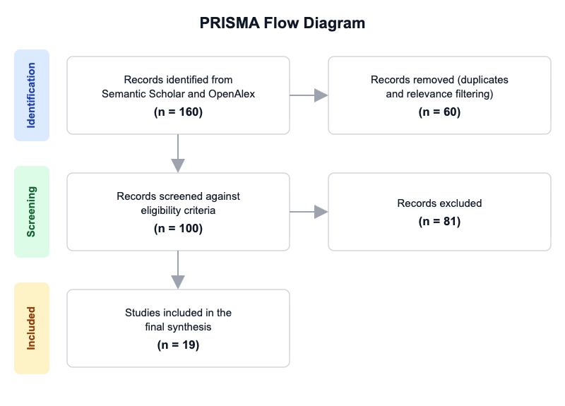
Initial database searching identified 160 records. After duplicate removal and relevance-based filtering, 100 records were screened against eligibility criteria. Of these, 81 papers were excluded, resulting in 19 papers included in the final synthesis.
PRISMA Flow Diagram
Eligibility criteria included:
- Human Study: Does the study involve human participants or human-derived clinical data rather than only animal or in vitro experiments?
- Dermatomyositis: Does the study focus on dermatomyositis or a clearly defined dermatomyositis subtype (juvenile, adult, amyopathic, or cancer-associated)?
- Cause or Treatment: Does the paper address either disease causes/pathogenesis or treatment/management of dermatomyositis?
- Clinical Relevance: Does the study report clinical outcomes, biomarkers, diagnostic features, prognosis, or treatment response relevant to dermatomyositis?
- Study Design: Is the article a clinical study, cohort, case series, guideline, review, or meta-analysis rather than a purely general commentary?
- Recent Evidence: Was the study published in 2020 or later?
All included studies met the stated eligibility criteria.
2.3 Data Extraction and Synthesis
Data extraction focused on the following variables:
- Cause/Mechanism: Extract the main proposed causes or pathophysiologic mechanisms discussed (e.g., autoantibodies, interferon pathway, complement-mediated vasculopathy, genetic associations, triggers).
- Patient Group: Extract the population studied or discussed (adult dermatomyositis, juvenile dermatomyositis, cancer-associated dermatomyositis, amyopathic dermatomyositis, mixed idiopathic inflammatory myopathies).
- Treatment: Extract the treatment, intervention, or management strategy evaluated or recommended (drug/class, biologic, IVIG, corticosteroids, steroid-sparing agent, nonpharmacologic management).
- Outcome: Extract the main clinical outcome(s) reported, including disease activity, muscle strength, rash improvement, remission, relapse, survival, or adverse events.
- Study Type: Extract the study design or article type (clinical trial, cohort, case series, review, meta-analysis, guideline, mechanistic study).
- Biomarker: Extract biomarkers, autoantibodies, imaging, histology, or laboratory markers linked to diagnosis, prognosis, or treatment response.
Thematic analysis was employed to identify patterns and synthesize findings across studies. Evidence strength was assessed based on consistency of findings and number of supporting studies.
3. Results
3.1 Characteristics of Included Studies
| Study | Year | Study Type | Population | Main Focus | Key Outcome / Finding |
|---|---|---|---|---|---|
| Cobos et al. [1] | 2020 | Review | Adult dermatomyositis | Diagnosis and treatment | Myositis-specific antibodies linked to phenotypes; therapies remain often refractory |
| Shirakashi et al. [2] | 2020 | Retrospective cohort | Anti-MDA5-positive DM-ILD | Plasma exchange in refractory RP-ILD | PE associated with survival benefit in treated intractable cases |
| Mehta et al. [3] | 2021 | Review | Anti-MDA5 DM, adult and juvenile; amyopathic forms | Pathophysiology, clinical features, management | RP-ILD drives survival; aggressive combined immunosuppression emphasized |
| Nombel et al. [4] | 2021 | Review | Anti-MDA5 dermatomyositis | Bioclinical features and emerging therapies | Interferon-mediated vasculopathy implicated; anti-MDA5 antibodies central |
| Leung et al. [5] | 2021 | Review | Juvenile dermatomyositis | Pathogenesis, assessment, management | Early aggressive therapy linked to better outcomes |
| Paik et al. [6] | 2022 | Systematic literature review | Adult DM and JDM | JAK inhibitors | Broad improvement in skin, muscle, and ILD reported |
| Kamperman et al. [7] | 2022 | Focused review | DM and IMNM | Pathophysiology and treatment | Conventional treatment often insufficient; new targets proposed |
| McCann et al. [8] | 2022 | Review | Juvenile dermatomyositis | Long-term outlook and emerging treatments | Distinct phenotypes with variable outlook and treatment needs |
| Tanboon & Nishino [9] | 2022 | Review | Adult dermatomyositis | Classification and biomarker-driven therapy | IFN1 pathway activation and DMSAs inform classification and treatment |
| Marzęcka et al. [10] | 2022 | Review | Adult dermatomyositis | Malignancy-associated autoantibodies | Anti-TIF1-γ, anti-NXP2, anti-SAE linked to cancer risk |
| Werth et al. [11] | 2022 | Phase 3 randomized trial | Adults with active DM | Lenabasum | Primary endpoint not met overall; subgroup signals seen |
| Lu et al. [12] | 2023 | Review | Anti-MDA5 DM | Pathogenesis and clinical progress | Viral trigger, immune-cell involvement, poor response to conventional therapy |
| Wang et al. [13] | 2023 | Systematic review and meta-analysis | Adult DM/PM with RP-ILD | Treatment comparisons | Biologics had highest remission; long-term prognosis poor |
| Cassard et al. [14] | 2024 | Review | Adult DM, including MDA5(+) and cancer-associated subtypes | Practical diagnosis and treatment guidance | Current treatment largely unchanged; targeted therapies needed |
| Castellini et al. [15] | 2024 | Systematic review and meta-analysis | DM and anti-MDA5 subsets | Type I IFN score | IFN score elevated in DM and anti-MDA5 disease |
| Coss et al. [16] | 2025 | Review | Juvenile dermatomyositis | Pathogenesis and therapy | IFN markers, SIGLEC-1, and CAR-T highlighted |
| Ali et al. [17] | 2025 | Review | Adult DM, classic and amyopathic | Cutaneous features, etiopathogenesis, treatment | Autoantibodies, IFNs, cytokines, and targeted therapies emphasized |
| Ng et al. [18] | 2025 | Review | Juvenile dermatomyositis | Pathogenesis and clinical applications | Biomarker-driven precision medicine and targeted therapies |
| Tripdatabase [19] | 2026 | Review | Adult dermatomyositis | Pathophysiology | Type I IFN dysregulation, autoantibodies, inflammatory/genetic mechanisms |
The evidence base is dominated by reviews and syntheses, with fewer primary treatment studies and one randomized phase 3 trial. Adult dermatomyositis, especially anti-MDA5-positive and malignancy-associated disease, is the most intensively studied subtype, while juvenile dermatomyositis is represented mainly by narrative reviews emphasizing pathogenesis and emerging therapies. Clinical outcomes range from biomarker-level disease activity to survival in rapidly progressive interstitial lung disease, making outcome comparability limited across designs.
3.2 Thematic Findings
3.2.1 Type I interferon dysregulation is the most consistent mechanistic axis across dermatomyositis subtypes
A convergent mechanistic signal across the literature is activation of the type I interferon pathway, which appears to unify classic adult dermatomyositis, anti-MDA5 disease, and juvenile dermatomyositis [9], [12], [16]. The strongest quantitative evidence comes from the meta-analysis showing higher type I IFN scores in dermatomyositis than healthy controls (pooled SMD = 2.27; 95% CI: 0.71, 3.82; p = 0.004) and in anti-MDA5-positive than anti-MDA5-negative disease (pooled SMD = 0.88; 95% CI: 0.06, 1.70; p = 0.03) [15]. Complementary review evidence links IFN pathway activation to dermatomyositis-specific antibodies, vasculopathy, and disease classification, with especially strong emphasis on interferon-related biomarkers such as IFN-regulated gene scores and SIGLEC-1 in juvenile disease [9], [16]. The same pathway is also used to explain why JAK inhibition, which interrupts IFN signaling, can improve skin, muscle, and lung manifestations [6]. Confidence: Strong.
3.2.2 Anti-MDA5 dermatomyositis represents a biologically severe phenotype marked by inflammatory and vascular injury
Anti-MDA5 dermatomyositis is repeatedly described as a distinct high-risk subtype characterized by rapidly progressive interstitial lung disease, distinctive skin findings, peripheral lymphopenia, and elevated serum ferritin [12], [4]. Mechanistic accounts converge on viral triggering in genetically predisposed hosts, activation of innate and adaptive immune cells, and interferon-mediated vasculopathy as central drivers of skin and lung injury [12], [4], [3]. Prognostic biomarker patterns further reinforce this biologic severity: higher neutrophil-lymphocyte ratio, serum ferritin, hypoxemia, HRCT score, rising KL-6, and cytokines including IL-1, IL-6, IL-8, IL-10, IL-12p70, IL-18, and sCD163 were associated with progression in intractable anti-MDA5-positive ILD [2]. The literature is highly consistent that this subset behaves differently from other dermatomyositis phenotypes, but the exact causal sequence remains unproven. Confidence: Strong.
3.2.3 Autoantibodies increasingly define diagnosis, prognosis, and cancer risk
Myositis-specific autoantibodies are no longer treated as ancillary laboratory findings; they now function as core stratification tools that link diagnosis to phenotype and prognosis [9], [14], [17]. Anti-MDA5 antibodies identify the severe lung-dominant phenotype, whereas anti-Mi-2, anti-TIF1γ, and anti-NXP2 help define other clinicopathologic subsets [9], [17]. In malignancy-associated dermatomyositis, anti-TIF1-γ was associated with a 9.37-fold higher cancer risk, anti-NXP2 with a 3.68-fold increase, and anti-SAE with malignancy in 14–57% of patients [10]. A further refinement is the observation that concurrent anti-CCAR1 and anti-TIF1-γ positivity identifies a lower-risk cancer-associated subgroup [9]. These findings support antibody-informed risk stratification, although the evidence is mostly observational and review-based. Confidence: Strong for phenotype association; Moderate for precise prognostic prediction.
3.2.4 Treatment is moving toward targeted rescue therapy, but conventional regimens still anchor care
Across the treatment literature, glucocorticoids and broad immunosuppressants remain the standard baseline approach, yet refractory disease is common and durable remission is inconsistent [7], [14], [17]. In anti-MDA5 disease, conventional glucocorticoid and immunosuppressant therapy is repeatedly described as poorly effective, prompting the use of aggressive combined immunosuppression, plasma exchange, and other rescue approaches [12], [2]. The strongest intervention signal comes from JAK inhibitors: among adult dermatomyositis cases, all 61 of 61 patients with refractory skin disease improved, all 16 of 16 with refractory muscle disease improved, and 31 of 33 with interstitial lung disease improved [6]. In contrast, lenabasum did not meet its primary endpoint overall in a phase 3 trial, with mean (SD) TIS 28.3 (19.75) vs 27.2 (19.23) at Week 28 for lenabasum 20 mg BID vs placebo and 40.6 (16.88) vs 34.8 (19.94) at Week 52; subgroup analyses suggested benefit in patients with muscle weakness and, separately, in those without muscle weakness for rash outcomes [11]. This pattern suggests that biologically targeted agents may work best when aligned with a specific inflammatory phenotype rather than the broader dermatomyositis label. Confidence: Moderate.
3.2.5 Juvenile dermatomyositis shows parallel but not identical biology to adult disease
Juvenile dermatomyositis is described as a systemic immune-mediated vasculopathy with a pronounced interferon signature, but the pediatric literature also emphasizes mitochondrial dysfunction, pyroptosis, neutrophil extracellular traps, and evolving B-cell biology [8], [18], [16]. Therapeutically, standard pediatric management still relies on high-dose corticosteroids with methotrexate, while newer agents such as JAK inhibitors, anti-IFN biologics, and CAR T-cell approaches are emerging [5], [16], [18]. The evidence base remains more developmental than definitive: reviews highlight favorable outcomes with early aggressive treatment and reports of drug-free remission, but high-quality comparative trials are still sparse [5], [16]. Although juvenile and adult disease share IFN biology, the pediatric literature suggests a broader translational pipeline and may serve as a testbed for precision immunotherapy. Confidence: Moderate.
3.2.6 Evidence for biologics and rescue therapies in interstitial lung disease suggests clinically meaningful benefit, but prognosis remains poor
Rapidly progressive interstitial lung disease is one of the most consequential complications in dermatomyositis, especially anti-MDA5-positive disease, and it strongly shapes survival [12], [4]. In the meta-analysis of adult DM/PM-RP-ILD, pooled prevalence was 8.9% (95% CI, 5.8% to 12.1%), remission rate was 58.4% (95% CI, 47.3% to 69.4%), and biologics had the highest remission and 6-month survival rates, whereas 1-year survival was 77.4% (95% CI, 66.7% to 88.1%) and 5-year survival was 40.0% (95% CI, 10.0% to 69.9%) [13]. Plasma exchange also showed a survival advantage in a refractory anti-MDA5-positive cohort, where 5 of 8 treated patients survived and 5 of 5 untreated patients died [2]. These findings align with the view that ILD is an inflammatory rescue window rather than a fully reversible complication, but long-term outcome remains guarded. Confidence: Moderate.
3.3 Summary of Evidence
| Theme | Key Finding | Population Applicability | Effect Direction | Confidence Level | Supporting Studies |
|---|---|---|---|---|---|
| Type I IFN activation | Type I IFN score higher in DM vs controls: pooled SMD = 2.27; 95% CI: 0.71, 3.82; p = 0.004 | Adult and mixed DM; partially matches juvenile disease | Positive | Strong | Castellini et al. [15], Tanboon & Nishino [9], Coss et al. [16] |
| Anti-MDA5 severe phenotype | Anti-MDA5 disease linked to RP-ILD, ferritin elevation, lymphopenia, and poor prognosis | Adult anti-MDA5 DM; partially matches exact DM population because it focuses on a subtype | Negative | Strong | Lu et al. [12], Nombel et al. [4], Mehta et al. [3] |
| Autoantibodies and cancer risk | Anti-TIF1-γ associated with 9.37-fold higher cancer risk; anti-NXP2 with 3.68-fold increase | Adult DM, especially malignancy-associated DM | Positive for risk detection | Strong | Marzęcka et al. [10], Cassard et al. [14], Cobos et al. [1] |
| JAK inhibitors | All 61/61 adult skin-refractory cases and 16/16 muscle-refractory cases improved; 31/33 adult ILD cases improved | Adult and juvenile DM; mixed but clinically relevant | Positive | Moderate | Paik et al. [6], Tanboon & Nishino [9], Coss et al. [16] |
| Conventional therapy limitations | Glucocorticoids and immunosuppressants often fail to yield sustained benefit | Adult DM and anti-MDA5 DM | Negative | Moderate | Kamperman et al. [7], Cassard et al. [14], Lu et al. [12] |
| Rescue therapy for RP-ILD | Plasma exchange: 5/8 survivors vs 5/5 deaths without PE in refractory anti-MDA5 ILD | Anti-MDA5 DM-ILD; subtype-specific and not general DM | Positive | Moderate | Shirakashi et al. [2], Wang et al. [13], Mehta et al. [3] |
| Juvenile dermatomyositis precision medicine | IFN scores, SIGLEC-1, and evolving targeted therapies support biomarker-driven care | Juvenile DM; does not fully match adult DM population | Positive | Moderate | Coss et al. [16], Ng et al. [18], Leung et al. [5] |
| Lenabasum trial | Primary endpoint not met: TIS 28.3 (19.75) vs 27.2 (19.23) at Week 28 | Adult DM with active skin disease | Null overall / mixed subgroup | Conflicting | Werth et al. [11], Cassard et al. [14], Ali et al. [17] |
4. Discussion
4.1 Principal Findings and Their Interpretation
The most important synthesis from the recent literature is that dermatomyositis is increasingly explained not as a single inflammatory syndrome but as a family of biologically patterned diseases organized around interferon activation, autoantibody-defined subgroups, and tissue-specific injury. The strength of the type I interferon signal is notable because it appears across different evidence layers: meta-analytic biomarker data, subtype-oriented reviews, and treatment logic all converge on the same pathway [15], [12], [16]. This convergence improves causal plausibility. If the same pathway is repeatedly elevated, linked to subtype severity, and therapeutically targeted by JAK inhibition, then the biomarker is more than a disease epiphenomenon; it becomes part of a coherent pathobiological model. That said, the literature still relies heavily on indirect inference rather than mechanistic human intervention studies, so causality remains supported but not proven.
The clearest example of biologic severity is anti-MDA5 dermatomyositis. Its association with rapidly progressive interstitial lung disease, ferritin elevation, lymphopenia, and macrophage-related markers suggests a highly inflammatory state in which vascular injury and innate immune activation dominate [12], [2]. This helps explain why conventional glucocorticoid-based therapy is often insufficient and why rescue strategies such as plasma exchange or aggressive combination immunosuppression are used [12], [2]. In other words, treatment response appears to depend on whether the dominant pathology is diffuse interferon-driven inflammation, severe pulmonary involvement, or chronic cutaneous disease.
The treatment signal is therefore strongest when therapy is matched to molecular phenotype. JAK inhibitors showed broad improvement across skin, muscle, and ILD manifestations in refractory disease [6], whereas lenabasum failed in the overall phase 3 population despite subgroup benefit [11]. This contrast likely reflects both biological heterogeneity and endpoint misalignment: a therapy that modulates inflammatory resolution may help selected symptom clusters but not the full mixed dermatomyositis population. Confidence is highest for interferon biology and anti-MDA5 severity, and lower for any single drug as a universal solution.
4.2 Comparison with Existing Literature and Resolution of Contradictions
The broad agreement between biomarker studies and clinical phenotype studies is meaningful because it reduces the likelihood that interferon activation is an isolated laboratory finding. Instead, it appears to track with actual disease expression, especially severe skin-lung disease and pediatric vasculopathy [15], [16], [12]. This consistency across adult and juvenile settings suggests that the interferon axis is a shared upstream driver, even if downstream manifestations differ by age, antibody profile, and organ involvement. The autoantibody literature reinforces this point by showing that anti-MDA5, anti-TIF1γ, and anti-NXP2 are not merely diagnostic labels but risk markers with different implications for lung disease and cancer [12], [10].
The main contradiction in the literature concerns treatment efficacy. JAK inhibition and plasma exchange appear promising, yet lenabasum did not meet its primary endpoint overall [6], [2], [11]. This is not necessarily a true refutation of targeted therapy; rather, it likely reflects patient heterogeneity, endpoint selection, and disease-domain mismatch. A drug directed toward inflammatory resolution may underperform in a trial mixing muscle-dominant, skin-dominant, and systemically severe phenotypes, especially when background immunosuppression is allowed. By contrast, observational reports in refractory and biomarker-enriched subgroups may overestimate benefit because they select patients already showing a strong interferon phenotype or imminent clinical deterioration. Publication bias is therefore plausible, particularly for novel agents and rescue interventions, because positive subgroup responses are more likely to be reported than null experiences. The most defensible interpretation is that dermatomyositis therapies are phenotype-dependent, and that negative broad-trial results do not invalidate targeted benefit in biologically enriched subsets.
4.3 Practical Implications
For clinicians, the evidence supports a subtype-first approach. Anti-MDA5-positive patients, and especially those with rapidly progressive interstitial lung disease, warrant urgent risk stratification using ferritin, lymphocyte counts, KL-6, HRCT severity, and cytokine-informed clinical assessment where available [2], [12]. In contrast, patients with chronic cutaneous disease may be better served by therapies targeting interferon signaling or skin inflammation, rather than escalation based solely on generalized immunosuppression [6], [11]. Pediatric care should also be more biomarker-informed, because juvenile disease shares interferon biology but differs in phenotype and long-term trajectory [8], [16].
For public health and specialty practice, the data argue for integrated autoantibody testing in suspected dermatomyositis because it informs both prognosis and downstream screening. Anti-TIF1γ, anti-NXP2, and anti-SAE positivity identifies higher malignancy risk and supports more selective cancer surveillance [10]. For regulatory and therapeutic development, the overall message is not that current standards should be abandoned, but that one-size-fits-all trial design is inadequate for dermatomyositis. Population-wide exposure reduction is not relevant here; instead, the analogous principle is phenotype-specific treatment selection and biomarker-enriched trial enrollment. This is especially important for anti-MDA5 disease, where conventional regimens often fail and rescue therapies may need earlier deployment [12], [2].
4.4 Strengths and Limitations
This review draws on a recent and diverse evidence base spanning mechanistic reviews, systematic reviews, meta-analyses, a cohort study, and a randomized phase 3 trial, which strengthens triangulation across biological and clinical domains. The synthesis is also strengthened by its focus on subtype-specific interpretation, which is essential in a heterogeneous disease like dermatomyositis.
The included literature nevertheless has important limitations. Many studies are narrative reviews rather than primary comparative studies, treatment data are often observational, and outcome measures vary widely across skin scores, muscle assessments, ILD endpoints, and survival. Juvenile and adult disease are frequently discussed together despite important biological differences, and several findings apply only to anti-MDA5 or malignancy-associated subgroups. For this review, the main limitation is that conclusions depend on the available extracted data and published abstracts, without a formal risk-of-bias assessment or full-text verification of every underlying study.
5. Gaps and Future Directions
The most pressing gap is the lack of prospective, biomarker-stratified trials that directly test whether interferon-high or anti-MDA5-positive patients benefit more from JAK inhibition, anti-IFN biologics, plasma exchange, or combination rescue therapy than unselected dermatomyositis populations. Current evidence repeatedly implies such heterogeneity, but does not yet prove optimal treatment allocation. Future studies should harmonize outcome definitions across muscle, skin, and lung domains, because inconsistent endpoints make cross-study comparison difficult and likely contributed to the mixed lenabasum findings. More work is also needed in juvenile dermatomyositis to connect IFN scores, SIGLEC-1, mitochondrial dysfunction markers, and CAR T-cell responses to durable clinical outcomes. The malignancy-risk literature would benefit from prospective validation of anti-TIF1γ, anti-NXP2, and anti-SAE for risk-adapted cancer screening. Finally, the field needs more primary mechanistic human studies linking autoantibodies, cytokines, and tissue pathology, since current causal claims remain inferential rather than experimentally confirmed.
6. Conclusion
The recent literature supports a clear but subtype-dependent answer: dermatomyositis is increasingly understood as an interferon-driven, autoantibody-patterned autoimmune disease, and treatment success depends on matching therapy to the underlying biologic phenotype. The strongest evidence points to type I interferon dysregulation as a central mechanism, with higher type I IFN scores in dermatomyositis than healthy controls (pooled SMD = 2.27; 95% CI: 0.71, 3.82; p = 0.004) and in anti-MDA5-positive versus anti-MDA5-negative disease (pooled SMD = 0.88; 95% CI: 0.06, 1.70; p = 0.03) [15]. Anti-MDA5 dermatomyositis stands out as the most severe subtype, especially when complicated by rapidly progressive interstitial lung disease, where ferritin, KL-6, HRCT severity, and cytokine markers help identify high-risk patients [2], [12]. On treatment, broad immunosuppression remains standard, but the most encouraging signals come from JAK inhibitors and selected rescue strategies such as plasma exchange, while lenabasum did not improve overall phase 3 outcomes despite subgroup signals [6], [2], [11]. The evidence base is strongest for mechanistic and prognostic stratification, but still only partly definitive for therapy choice because many studies are subtype-specific, observational, or review-based. The single most important unresolved question is which biomarker-defined patients should receive which targeted therapy first. Answering that question will be crucial for improving survival, reducing toxicity, and making dermatomyositis care truly precision-based.
References
- Cobos, G. A., Femia, A. N., & Vleugels, R. A. (2020). Dermatomyositis: An update on diagnosis and treatment. American Journal of Clinical Dermatology, 21(3), 339–353. https://doi.org/10.1007/s40257-020-00502-6
- Shirakashi, M., Nakashima, R., Tsuji, H., Tanizawa, K., Handa, T., Hosono, Y., Akizuki, S., Murakami, K., Hashimoto, M., Yoshifuji, H., Ohmura, K., & Mimori, T. (2020). Efficacy of plasma exchange in anti-MDA5-positive dermatomyositis with interstitial lung disease under combined immunosuppressive treatment. Rheumatology, 59(11), 3284–3292. https://doi.org/10.1093/rheumatology/keaa123
- Mehta, P., Machado, P. M., & Gupta, L. (2021). Understanding and managing anti-MDA 5 dermatomyositis, including potential COVID-19 mimicry. Rheumatology International, 41(6), 1021–1036. https://doi.org/10.1007/s00296-021-04819-1
- Nombel, A., Fabien, N., & Coutant, F. (2021). Dermatomyositis with anti-MDA5 antibodies: Bioclinical features, pathogenesis and emerging therapies. Frontiers in Immunology, 12, 773352. https://doi.org/10.3389/fimmu.2021.773352
- Leung, A. K. C., Lam, J. M., Alobaida, S., Leong, K. F., & Wong, A. H. C. (2021). Juvenile dermatomyositis: Advances in pathogenesis, assessment, and management. Current Pediatric Reviews, 17(4), 273–287. https://doi.org/10.2174/1573396317666210426105045
- Paik, J. J., Lubin, G., Gromatzky, A., Mudd, P. N., Jr., Ponda, M. P., & Christopher-Stine, L. (2022). Use of Janus kinase inhibitors in dermatomyositis: A systematic literature review. Clinical and Experimental Rheumatology, 41(2), 348–358. https://doi.org/10.55563/clinexprheumatol/hxin6o
- Kamperman, R. G., van der Kooi, A. J., de Visser, M., Aronica, E., & Raaphorst, J. (2022). Pathophysiological mechanisms and treatment of dermatomyositis and immune mediated necrotizing myopathies: A focused review. International Journal of Molecular Sciences, 23(8), 4301. https://doi.org/10.3390/ijms23084301
- McCann, L. J., Livermore, P., Wilkinson, M. G. L., & Wedderburn, L. R. (2022). Juvenile dermatomyositis. Where are we now? Clinical and Experimental Rheumatology, 40(2), 394–403. https://doi.org/10.55563/clinexprheumatol/56ilob
- Tanboon, J., & Nishino, I. (2022). Update on dermatomyositis. Current Opinion in Neurology, 35(5), 611–621. https://doi.org/10.1097/WCO.0000000000001091
- Marzęcka, M., Niemczyk, A., & Rudnicka, L. (2022). Autoantibody markers of increased risk of malignancy in patients with dermatomyositis. Clinical Reviews in Allergy & Immunology, 63(2), 289–296. https://doi.org/10.1007/s12016-022-08922-4
- Werth, V. P., White, B., Dgetluck, N., Hally, K., Constantine, S., Aggarwal, R., Fiorentino, D. F., Lundberg, I. E., & Oddis, C. V. (2022). OP0162 Efficacy and safety of lenabasum in the phase 3 DETERMINE trial in dermatomyositis [Conference abstract]. Annals of the Rheumatic Diseases, 81(Suppl 1), 106–107. https://doi.org/10.1136/annrheumdis-2022-eular.5037
- Lu, X., Peng, Q., & Wang, G. (2023). Anti-MDA5 antibody-positive dermatomyositis: Pathogenesis and clinical progress. Nature Reviews Rheumatology, 20(1), 48–62. https://doi.org/10.1038/s41584-023-01054-9
- Wang, H., Lv, J., He, J., Wu, W., Zhong, Y., Cao, S., Cai, Y., & Wang, Q. (2023). The prevalence and effects of treatments of rapidly progressive interstitial lung disease of dermatomyositis/polymyositis adults: A systematic review and meta-analysis. Autoimmunity Reviews, 22(8), 103335. https://doi.org/10.1016/j.autrev.2023.103335
- Cassard, L., Seraly, N., Riegert, M., Patel, A., & Fernandez, A. P. (2024). Dermatomyositis: Practical guidance and unmet needs. ImmunoTargets and Therapy, 13, 151–172. https://doi.org/10.2147/ITT.S381472
- Castellini, C., Scotti, C., Navarini, L., Fu, Q., Qian, J., Giacomelli, R., Cavagna, L., & Ruscitti, P. (2024). The evaluation of type I interferon score in dermatomyositis, a systematic review and a meta-analysis. Autoimmunity Reviews, 23(12), 103686. https://doi.org/10.1016/j.autrev.2024.103686
- Coss, S. L., Sabbagh, S. E., & Kim, H. (2025). Updates in juvenile dermatomyositis: Pathogenesis and therapy. Current Opinion in Rheumatology, 37(6), 445–456. https://doi.org/10.1097/BOR.0000000000001112
- Ali, H., On, A., Xing, E., Shen, C., & Werth, V. P. (2025). Dermatomyositis: Focus on cutaneous features, etiopathogenetic mechanisms and their implications for treatment. Seminars in Immunopathology, 47(1), 32. https://doi.org/10.1007/s00281-025-01054-9
- Ng, P. P. L. C., Syntakas, A. E., & Wedderburn, L. R. (2025). Juvenile dermatomyositis: New insights into pathogenesis and clinical applications. Pediatric Rheumatology, 23(1), 115. https://doi.org/10.1186/s12969-025-01146-8
- Trip Database. (2026). Dermatomyositis [Evidence database record]. https://www.tripdatabase.com/