

Latest Research on Invasive Lobular Carcinoma (ILC): A Thematic Literature Review of Diagnosis, Genomics, Prognosis, and Treatment
Abstract
Recent research on invasive lobular carcinoma (ILC) converges on a consistent view that this is a biologically and clinically distinct breast cancer subtype, defined by E-cadherin/CDH1 disruption, enriched PI3K-pathway alterations, endocrine sensitivity, and delayed but persistent relapse risk. Across genomic cohorts, CDH1 alteration rates reached 65% in primary disease and 76%-79% in metastatic cohorts, while PIK3CA alterations were present in 48%-58% of metastatic cases and PTEN alterations in 6%-11%; in metastatic ILC, overall survival differed by CDH1 mutation status (5.3 years wild-type vs 3.7 years mutated, p=0.01), PIK3CA status (4.6 years vs 3.4 years, p<0.001), and PTEN status (4.2 years vs 3.4 years, p=0.008) [16], [17]. Clinically, ILC shows inferior pathologic response to chemotherapy but better endocrine-therapy-associated outcomes, including 10-year distant recurrence-free survival of 65% with neoadjuvant endocrine therapy versus 55% with neoadjuvant chemotherapy (HR 0.57, 95% CI 0.33-0.98, P = 0.02) [14]. Diagnostic and prognostic advances now include standardized E-cadherin immunohistochemistry, which improves interpathologist agreement, and mPEPI-based risk stratification, where recurrence-free survival at 100 months was 82.2% for mPEPI 0-1 versus 73.6% for mPEPI 2-3 (p=0.04) [11], [15]. Despite progress, ILC remains underrepresented in trials, limiting certainty about optimal systemic therapy, especially in metastatic settings and in CDH1-negative edge cases [13], [12].
1. Introduction
Invasive lobular carcinoma (ILC) accounts for roughly 10%-15% of breast cancers and is the second most common invasive subtype, yet it remains less well characterized than invasive ductal or no-special-type disease. Its clinical importance is disproportionate to its frequency because its biology differs in fundamental ways: loss of E-cadherin function drives a discohesive, single-file growth pattern; tumors are often estrogen receptor-positive, HER2-negative, and clinically difficult to detect because of multifocality, multicentricity, and subtle imaging appearance [8], [10], [6]. These features are not merely descriptive. They affect staging, surgical planning, response to systemic therapy, and the timing of recurrence, with multiple reports indicating that ILC behaves as a late-relapsing disease with metastatic predilection for sites such as bone, serosa, meninges, ovaries, and liver [4], [6], [7].
The research landscape has expanded rapidly over the last decade, moving from broad clinicopathologic comparison toward more granular genomic, translational, and treatment-focused work. This shift is important because ILC has historically been folded into studies of more common breast cancer histologies, limiting the ability to infer subtype-specific prognosis or treatment benefit. Recent studies increasingly address whether standard chemotherapy, endocrine therapy, and newer targeted approaches perform differently in ILC, while parallel diagnostic research examines how to improve pathological classification and prognostic stratification [14], [11], [15]. At the same time, molecular studies have clarified that beyond CDH1 loss, ILC is enriched for alterations in PIK3CA, PTEN, AKT1, FOXA1, ERBB2, NF1, RB1, GATA3, AXIN2, and CTNND1, suggesting several biologically meaningful subgroups with potential therapeutic implications [1], [2], [12], [16], [18].
This review synthesizes the latest evidence on ILC as it relates to diagnosis, molecular mechanisms, prognosis, treatment response, and translational opportunities, with particular emphasis on what has changed in the most recent literature and where uncertainty remains most consequential for clinical care.
2. Methods
2.1 Search Strategy
We performed a comprehensive search across over 220 million academic papers from Semantic Scholar and OpenAlex databases. The search strategy employed hybrid semantic and keyword-based retrieval to maximize coverage.
Search queries included:
- "Invasive lobular carcinoma latest research diagnosis prognosis therapy"
- "Invasive lobular breast cancer molecular biology genomic alterations"
- "Invasive lobular carcinoma treatment outcomes endocrine therapy metastasis"
- "Systematic review invasive lobular carcinoma recent advances"
- "Metastatic invasive lobular carcinoma clinical studies recent"
2.2 Study Selection
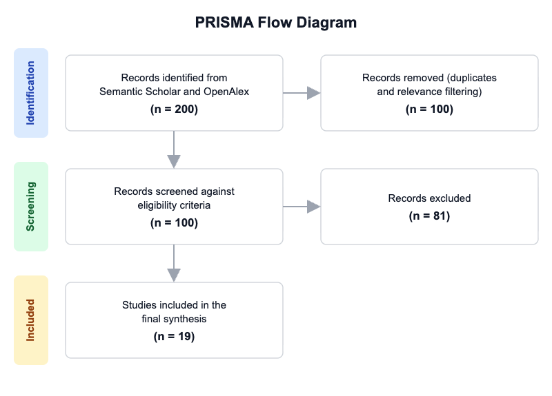
Initial database searching identified 200 records. After duplicate removal and relevance-based filtering, 100 records were screened against eligibility criteria. Of these, 81 papers were excluded, resulting in 19 papers included in the final synthesis.
PRISMA Flow Diagram
Eligibility criteria included:
- ILC Focus: Does the study primarily concern invasive lobular carcinoma or lobular breast cancer in humans, patient samples, or validated disease models?
- Recent Evidence: Does the study report original evidence or a synthesis published in the last 10 years, rather than only older foundational work?
- Clinical or Translational: Does the study provide clinically relevant or translational information on diagnosis, prognosis, treatment, metastasis, pathology, or biomarkers in ILC?
- Human or Validated Model: Does the study use human participants, human tumor samples, organoids, cell lines, animal models, or computational analyses relevant to ILC?
- Molecular Insight: Does the study report a molecular, genomic, histologic, imaging, or mechanistic insight relevant to ILC biology?
- Therapeutic Insight: Does the study evaluate treatment response, therapeutic resistance, or actionable targets in ILC?
- Diagnostic or Prognostic Insight: Does the study evaluate diagnosis, screening, staging, risk stratification, or survival/prognostic outcomes in ILC?
All included studies met the stated eligibility criteria.
2.3 Data Extraction and Synthesis
Data extraction focused on the following variables:
- Research Focus: Identify the main ILC topic addressed (e.g., diagnosis, prognosis, molecular biology, metastasis, treatment, imaging, pathology, biomarkers).
- Study Type: Extract the study design or evidence type (clinical trial, cohort, case-control, review, systematic review, meta-analysis, translational study, genomic analysis).
- Cohort/Model: Specify the patient population, tumor subtype, cell line, animal model, or dataset used, including any notable inclusion criteria if stated.
- Key Finding: Summarize the principal ILC-related finding or conclusion stated in the abstract or results.
- Biomarker/Target: Extract any molecular marker, gene, pathway, receptor, or therapeutic target highlighted as important in the paper.
- Clinical Relevance: Describe the stated or implied relevance to ILC diagnosis, risk stratification, prognosis, treatment selection, or disease monitoring.
- Methods/Platform: Extract the main method, assay, imaging modality, sequencing platform, or analytical approach used.
- Limitations/Future Work: Capture any limitations, gaps, or future research directions explicitly mentioned by the authors.
Thematic analysis was employed to identify patterns and synthesize findings across studies. Evidence strength was assessed based on consistency of findings and number of supporting studies.
3. Results
3.1 Characteristics of Included Studies
| Study and Year | Study Type | Population / Cohort | Key Focus | Main Method | Primary Outcome |
|---|---|---|---|---|---|
| Desmedt et al. 2016 [1] | Genomic analysis | 630 primary ILC tumors | Genomic characterization | Sequencing and copy-number analysis | Altered CDH1, PIK3CA, PTEN, AKT1, HER2/HER3, FOXA1, ESR1 |
| Ciriello et al. 2015 [2] | Multidimensional molecular profiling | 817 breast tumors including 127 ILC | Molecular portraits | Integrated genomic profiling | ILC-enriched PTEN, TBX3, FOXA1 alterations and transcriptional subtypes |
| Cristofanilli et al. 2004 [3] | Cohort analysis | 1,034 stage II-III breast cancers; 122 ILC | Chemotherapy response | Clinical trial cohort analysis | Lower pCR but longer recurrence-free and overall survival in ILC |
| Pestalozzi et al. 2008 [4] | Retrospective cohort | 12,206 trial participants; 9,374 pure IDC/ILC | Prognosis and recurrence | Trial data synthesis | Distinct recurrence pattern and survival timing in ILC |
| McCart Reed et al. 2021 [5] | Review | Not specified | Pathology, genomics, management | Narrative review | Distinct biology, endocrine sensitivity, late recurrence |
| Pramod et al. 2021 [6] | Comprehensive review | Not specified | Molecular mechanisms and clinical features | Literature synthesis | E-cadherin loss, FOXA1 and GATA3 differences vs IDC |
| Mouabbi et al. 2022 [7] | Review | Not specified | Diagnostic and treatment strategy | Narrative review | Need for dedicated ILC trials |
| Van Baelen et al. 2022 [8] | Review | Not specified | Diagnostic and future treatment strategies | Literature synthesis | ILC as distinct malignancy with diagnostic and therapeutic differences |
| Thomas et al. 2019 [9] | Review | Not specified | Early-stage management | Literature review | Sparse treatment-specific evidence for ILC |
| Batra et al. 2023 [10] | Narrative review | Not specified | Pathology and translational features | Review with technological synthesis | Endocrine therapy, CDK4/6 inhibitors, spatial transcriptomics |
| De Schepper et al. 2024 [11] | Translational diagnostic study | E-cadherin antibody testing in ILC-related lesions | Diagnostic standardization | IHC and Western blot | Improved interpathologist agreement |
| Dopeso et al. 2024 [12] | Genomic and epigenomic analysis | 364 primary ILCs | CDH1-negative ILC biology | Targeted sequencing and WGS | AXIN2 and CTNND1 as alternative drivers |
| Van Baelen et al. 2024 [13] | Systematic review | 93 phase III/IV trials | Trial representation | PRISMA-guided review | ILC poorly documented and underrepresented |
| Mouabbi et al. 2025 [14] | Retrospective cohort | 611 stage II-III ER+ HER2- ILC patients | NAET vs NAC | Institutional database analysis | NAET associated with superior 10-year DRFS |
| Vertido et al. 2026 [15] | Retrospective prognostic study | 124 ER+ HER2- ILC patients treated with NET | mPEPI validation | Kaplan-Meier analysis | mPEPI stratified recurrence risk |
| Shen et al. 2025 [16] | Retrospective cohort | 654 metastatic ILC patients | Treatment patterns and genomics | MSK-IMPACT NGS | OS associated with CDH1, PIK3CA, PTEN |
| Fanucci et al. 2026 [17] | Genomic cohort study | 533 metastatic ER+/HER2- patients; 125 ILC | Metastatic genomics vs NST | OncoPanel sequencing | Distinct ILC genomic landscape, similar outcomes to NST |
| Farrokhi and Stenehjem 2026 [18] | Observational real-world study | 256 metastatic HR+ HER2- ILC patients | Alteration-specific outcomes | Clinicogenomics database | NF1, RB1, GATA3, ARID1A linked to poorer outcomes |
| Lin and Mesias 2024 [19] | Population-based retrospective study | 343,397 IDC and 39,859 ILC | Short- and long-term survival | SEER analysis | Early BCSS advantage but later disadvantage in ILC |
Across the included literature, the evidence base spans pathology, genomic profiling, retrospective clinical cohorts, population-level survival analyses, and multiple narrative or systematic reviews. The dominant clinical populations are hormone receptor-positive, HER2-negative ILC in both early-stage and metastatic settings, while diagnostic studies focus on E-cadherin interpretation and pathology standardization. The largest and most consistent body of primary evidence concerns genomic hallmarks and treatment resistance, whereas trial-design representation and prognostic tool validation remain comparatively sparse.
3.2 Thematic Findings
3.2.1 ILC is a molecularly defined subtype centered on cell-adhesion loss with recurrent pathway-level alterations
The most consistent mechanistic finding across studies is that ILC is anchored by disruption of cell adhesion, especially CDH1/E-cadherin loss, but is not molecularly reducible to CDH1 alone. In primary disease, CDH1 mutation was present in 65% of tumors, while alterations in PIK3CA, PTEN, and AKT1 each appeared in more than one-half of cases in combined form across the PI3K axis, and HER2/HER3, FOXA1, and ESR1 alterations were also recurrent [1]. In metastatic ILC, CDH1 mutations remained highly prevalent at 79%, with PIK3CA at 48%, PTEN at 11%, ESR1 at 9.3%, HER2 at 17%, AKT1 at 5.6%, and NF1 at 6% [16]. A separate metastatic cohort similarly identified CDH1 (76%) and PIK3CA (58%) as dominant events, with TP53, ESR1, PTEN, NF1, MAP3K1, ERBB2, and AKT1 also recurrent [17].
What emerges from these data is a convergent model in which loss of adhesion coexists with altered PI3K/ERBB/endocrine signaling, offering a plausible biological basis for ILC’s discohesive growth and therapy resistance. The finding that ILCs lacking CDH1 bi-allelic inactivation may instead harbor AXIN2 or CTNND1 alterations, and that AXIN2 knockout induces lobular-like migration and anoikis resistance, further supports a shared cell-adhesion phenotype rather than a single-gene explanation [12]. Confidence: Strong.
3.2.2 Prognosis in ILC is time-dependent, with early apparent benefit followed by late relapse risk
Survival patterns in ILC are consistently non-linear over time, which helps reconcile apparently conflicting short- and long-term results across cohorts. In trial-based analyses, ILC showed an early advantage in disease-free and overall survival that reversed after 6 and 10 years, respectively [4]. In population-level analysis, breast cancer-specific survival was better in ILC during the first five years (HR=0.71, p<0.001) but worse thereafter, with HR=1.30 in years 6-10, HR=1.75 in years 11-15, and HR=2.17 in years 16-20 [19]. Similarly, older chemotherapy cohort data found longer recurrence-free survival and overall survival in ILC despite lower pathologic complete response, suggesting that pCR may be a weak surrogate in this subtype [3].
This time-dependent pattern implies that early treatment metrics can be misleading in ILC because the disease’s low-proliferative, endocrine-responsive biology may delay recurrence rather than prevent it. The consistency of late hazard acceleration across datasets makes late relapse a robust clinical feature rather than an artifact of a single cohort. Confidence: Strong.
3.2.3 Endocrine sensitivity is real, but it does not fully eliminate recurrence risk
Several studies converge on the notion that ILC is strongly endocrine responsive, yet endocrine sensitivity coexists with persistent residual risk. Most ILC tumors are estrogen receptor-positive and HER2-negative, and the biology is often described as luminal [7], [10], [6]. In stage II-III ER+ HER2- ILC, neoadjuvant endocrine therapy achieved a 10-year distant recurrence-free survival of 65% versus 55% for neoadjuvant chemotherapy (HR 0.57, 95% CI 0.33-0.98, P = 0.02), while pathologic complete response remained very low in both arms: 2.5% with chemotherapy and 1.9% with endocrine therapy [14]. In a separate NET-treated cohort, mPEPI 0-1 was associated with 100-month recurrence-free survival of 82.2% versus 73.6% for mPEPI 2-3 (p=0.04) [15].
Together, these data indicate that endocrine treatment can be effective for many patients with ILC, but classical response endpoints such as pCR may underestimate benefit, and low-risk post-treatment profiles still leave substantial recurrence risk. The clinical implication is not that chemotherapy is useless in all settings, but that endocrine-based strategies likely fit the subtype’s low-proliferative biology better than cytotoxic approaches in many early-stage cases. Confidence: Moderate to strong.
3.2.4 Chemotherapy resistance is a recurring clinical feature, but its prognostic meaning differs by setting
ILC repeatedly shows limited pathologic response to chemotherapy. In the primary chemotherapy cohort, pCR was 3% in ILC versus 15% in IDC [3]. In the neoadjuvant comparison of stage II-III ER+ HER2- ILC, pCR remained similarly low with 2.5% for NAC and 1.9% for NAET [14]. Yet low pCR does not translate into uniformly poor long-term outcomes, because the same chemotherapy cohort showed better recurrence-free and overall survival for ILC than IDC despite inferior pathologic response [3].
This is an important contradiction only on the surface. The likely explanation, supported by the survival data, is that ILC is less chemosensitive in the short term but may be biologically more indolent, so pCR is not calibrated to capture its prognostic trajectory. The evidence therefore argues against using chemotherapy response alone to judge ILC prognosis. Confidence: Strong.
3.2.5 Genomic alterations increasingly appear clinically actionable in metastatic ILC, but outcome associations are alteration-specific
Metastatic ILC is not genomically uniform. In one large single-center cohort, OS differed by CDH1 mutation status (5.3 years wild-type vs 3.7 years mutated, p=0.01), PIK3CA status (4.6 years vs 3.4 years, p<0.001), and PTEN status (4.2 years vs 3.4 years, p=0.008), while HER2, AKT1, and ESR1 status were not associated with OS [16]. In a separate real-world cohort, NF1 alterations were associated with worse time to next treatment and time to treatment discontinuation, RB1 alterations with shorter time to next treatment and treatment discontinuation, ARID1A with shorter overall survival, and GATA3 with shorter treatment discontinuation in the CDK4/6 inhibitor plus endocrine therapy subgroup [18]. Another metastatic sequencing study found CDH1 and ERBB2 alterations associated with early recurrence, and ERBB2 alterations enriched among patients with liver metastases [17].
The synthesis suggests that metastatic ILC is best understood as a pathway-diverse disease in which some alterations track with endocrine resistance, some with broader poor prognosis, and some with organotropic spread. However, not every recurrent alteration has proven prognostic value, and null results for HER2, AKT1, and ESR1 in one cohort illustrate that prevalence alone does not imply outcome impact. Confidence: Moderate.
3.2.6 Diagnosis and trial design remain major bottlenecks, despite advances in pathology standardization
Diagnostic work now reinforces that pathology can be standardized more effectively, but representation in clinical research remains inadequate. E-cadherin immunohistochemistry improved interpathologist agreement for ILC and related lobular lesions, and antibody clone performance varied by biochemical specificity, with reduced specificity reported for EP700Y and NCL-L-E-cad and cross-reactivity for Clone 36 to P-cadherin [11]. At the trial level, ILC was poorly documented in phase III/IV studies; inclusion rates ranged from 2.0% to 26.0%, and central pathology was not reported [13].
This combination is consequential: diagnostic reliability is improving within pathology practice, but evidence generation still lags because ILC is systematically underrepresented in treatment trials. As a result, clinical decisions are often extrapolated from broader breast cancer populations whose biology only partially matches ILC. Confidence: Strong for diagnostic standardization; moderate for the broader trial-representation implication.
3.3 Summary of Evidence
| Theme | Key Finding | Population Applicability | Effect Direction | Confidence Level | Supporting Studies |
|---|---|---|---|---|---|
| Adhesion-loss and pathway-level genomics define ILC | CDH1 mutation 65% in primary ILC; 79% in metastatic ILC; PIK3CA 48%-58%, PTEN 6%-11%, ESR1 9.3%-10%, and NF1 6%-16% recurrent | Primary and metastatic ILC; mostly HR+/HER2- metastatic cohorts | Mixed, but consistently ILC-defining | Strong | Desmedt et al. [1], Shen et al. [16], Fanucci et al. [17] |
| Time-dependent prognosis with late relapse | ILC had better early BCSS (HR=0.71) but worse later BCSS (HR=1.30; HR=1.75; HR=2.17 over later intervals) | Stage I-III ILC, including population-based comparisons with IDC | Mixed | Strong | Lin and Mesias [19], Pestalozzi et al. [4], Cristofanilli et al. [3] |
| Endocrine responsiveness and prognostic tools | NAET vs NAC DRFS 65% vs 55% (HR 0.57, 95% CI 0.33-0.98); mPEPI 0-1 vs 2-3 RFS 82.2% vs 73.6% | ER+, HER2- early-stage ILC treated with endocrine-based neoadjuvant therapy | Positive for endocrine-based approaches | Moderate to strong | Mouabbi et al. [14], Vertido et al. [15] |
| Chemotherapy resistance with limited pCR utility | pCR 3% in ILC vs 15% in IDC; NAC pCR 2.5% vs NAET 1.9% in stage II-III ILC | Stage II-III ILC, compared with IDC or within ILC neoadjuvant cohorts | Negative for pCR as response metric; mixed for prognosis | Strong | Cristofanilli et al. [3], Mouabbi et al. [14] |
| Metastatic genomics and outcome stratification | OS differed by CDH1 (5.3 vs 3.7 years), PIK3CA (4.6 vs 3.4 years), PTEN (4.2 vs 3.4 years), NF1 altered TTNT/TTD, RB1 altered TTNT/TTD, ARID1A shortened OS | Metastatic ILC, mostly HR+/HER2- | Negative for certain alterations, null for others | Moderate | Shen et al. [16], Farrokhi and Stenehjem [18], Fanucci et al. [17] |
| Diagnostic standardization and trial underrepresentation | E-cadherin IHC improved interpathologist agreement; ILC documented in only 14% of trial manuscripts with inclusion rates 2.0%-26.0% | Diagnostic pathology and phase III/IV trial populations | Positive for diagnostic reliability; negative for trial representation | Strong | De Schepper et al. [11], Van Baelen et al. [13] |
4. Discussion
4.1 Principal Findings and Their Interpretation
The central message of the contemporary ILC literature is that this disease is not simply a histologic variant of breast cancer but a biologically coherent subtype with recognizable molecular dependencies and a clinically distinctive natural history. The strongest mechanistic anchor is adhesion loss, usually via CDH1 inactivation, but the newer work makes clear that the lobular phenotype can also arise through alternative cell-adhesion lesions such as AXIN2 and CTNND1 disruption [12]. This is important because it suggests that the invasive pattern of ILC reflects convergent biology rather than a single mutational event. The recurrent co-alteration of PI3K-pathway genes such as PIK3CA, PTEN, and AKT1, together with ERBB2/ERBB3 and FOXA1, provides a plausible pathway-level explanation for both endocrine sensitivity and treatment escape [1], [2], [17]. In other words, the clinical phenotype appears to be driven by a combination of low-adhesion growth and endocrine-pathway rewiring.
The time-dependent survival pattern is equally informative. Better early outcomes, followed by late hazard accumulation, imply that ILC often behaves as a slow-burn disease rather than an acutely aggressive one. That pattern fits the observed high ER positivity, lower grade, and reduced chemosensitivity, while also explaining why short-term response metrics are a poor summary of true benefit [4], [19], [3]. The neoadjuvant endocrine data strengthen this interpretation: endocrine therapy appears better aligned to ILC biology than chemotherapy, but the absolute recurrence risk remains meaningful, which is why mPEPI and related post-treatment biomarkers matter [14], [15]. Confidence is highest for the existence of endocrine sensitivity and late relapse risk, and lower for any single genomic marker beyond CDH1, PIK3CA, and PTEN because alteration-specific outcome data remain context dependent.
4.2 Comparison with Existing Literature and Resolution of Contradictions
The apparent contradiction between low chemotherapy response and sometimes favorable long-term survival is resolved when ILC is viewed through its proliferative and endocrine context. Chemotherapy response assays such as pCR are optimized for tumors in which rapid cytotoxic kill tracks closely with prognosis; ILC does not appear to fit that model well [3]. The better early survival in ILC, followed by late deterioration, is consistent with an indolent disease that escapes early detection and later manifests as delayed relapse, rather than one in which immediate chemosensitivity determines outcome [4], [19]. This means the seeming mismatch between response and prognosis is not truly contradictory; it reflects a biomarker-prognosis mismatch.
There is more genuine heterogeneity in the metastatic genomics literature. Some studies report prognostic associations for CDH1, PIK3CA, PTEN, NF1, RB1, ARID1A, and GATA3, whereas others find no OS effect for HER2, AKT1, or ESR1 despite clear recurrence [16], [18], [17]. The most evidence-supported explanation is that outcome associations differ by endpoint, treatment line, and disease context: some alterations likely influence early endocrine escape, whereas others mark broader genomic instability or metastatic aggressiveness. The retrospective and single-center nature of much of the evidence also raises the possibility of selection bias and confounding by therapy sequencing. Publication bias is plausible as well, because positive genomic associations are more likely to be reported than null findings, especially in a disease where sample sizes are small relative to heterogeneity. The most conservative interpretation is therefore that metastatic ILC is genomically actionable, but the actionable subset is not yet fully defined.
Methodologically, the field has improved from broad IDC-vs-ILC comparisons toward subtype-specific sequencing and pathology harmonization. That progression increases confidence in the direction of the evidence, even if it does not yet eliminate uncertainty about which alterations should change routine treatment.
4.3 Practical Implications
For clinicians, the immediate implication is that ILC should not be managed as a generic hormone receptor-positive breast cancer without subtype awareness. Endocrine-based neoadjuvant strategies appear particularly relevant in stage II-III ER+ HER2- disease, whereas pCR should be interpreted cautiously because it is a weak response surrogate in ILC [14], [3]. In practice, post-treatment risk tools such as mPEPI may help distinguish patients with meaningfully lower recurrence risk from those who remain vulnerable despite endocrine responsiveness [15].
For pathologists, routine E-cadherin immunohistochemistry, used with standardized clone selection and careful interpretation, can improve classification consistency and reduce diagnostic ambiguity in lobular lesions [11]. This matters because undercalling or overcalling lobular disease can distort treatment planning, imaging expectations, and trial eligibility.
For oncology teams treating metastatic disease, genomic profiling is increasingly justified because CDH1, PIK3CA, PTEN, NF1, RB1, and ARID1A each carry distinct prognostic or resistance implications in different cohorts [16], [18], [17]. However, the evidence does not yet support a single universal genomic rule for all metastatic ILC. The practical approach is therefore risk-adapted and alteration-informed rather than mutation-deterministic.
At the policy level, the trial literature indicates that ILC remains underrepresented and often poorly documented, which weakens external validity for this subtype [13]. That gap argues for histology-specific trial reporting and eligibility frameworks that recognize ILC’s non-measurable infiltrative behavior rather than relying on criteria designed for more circumscribed tumors.
4.4 Strengths and Limitations
A major strength of this review is the integration of pathology, genomics, prognostic modeling, and treatment-response evidence into a single subtype-focused synthesis. The included literature spans diagnostic standardization, primary and metastatic molecular profiling, population-based survival, and comparative neoadjuvant outcome studies, allowing the review to connect mechanistic biology with clinical consequence.
The limitations of the included studies are substantial and shape confidence. Much of the primary evidence is retrospective, single-center, or database-based, and populations are frequently enriched for hormone receptor-positive, HER2-negative disease, which only partially captures the full ILC spectrum. Several studies rely on surrogate outcomes such as pCR, time to treatment discontinuation, or recurrence-free survival, which are informative but not equivalent. The review-level evidence also includes narrative syntheses and systematic review of trial reporting rather than randomized ILC-specific trials.
This review is itself limited by abstract-based extraction for the source set and by the absence of a formal risk-of-bias assessment. Although the synthesis is comprehensive within the provided material, some potentially important details, such as assay-level validation metrics or subgroup-level confounding control, are not fully reported in the source abstracts.
5. Gaps and Future Directions
The most important gap is the lack of prospective, ILC-specific trials that test treatment strategies against endpoints meaningful for this subtype. Current evidence suggests endocrine-based neoadjuvant therapy is promising, but confirmation in larger, prospectively designed ILC cohorts is still needed, particularly for patients with stage-specific and menopausal subgroup differences [14], [15]. Another unresolved issue is how to classify and manage CDH1-negative lobular tumors, because AXIN2 and CTNND1 alterations appear to converge on the same phenotype, but these cases remain rare and incompletely characterized [12].
Methodologically, future studies should harmonize outcome definitions across early and metastatic disease, especially for late relapse, treatment discontinuation, and endocrine resistance. Genomic studies also need clearer separation of prognostic versus predictive effects, since some alterations appear to influence survival, whereas others may only mark disease burden or treatment history. The trial literature suggests a further need for ILC-aware eligibility criteria and reporting standards, because underrepresentation in phase III/IV studies limits generalizability [13]. Finally, future research should expand beyond ER+/HER2- disease to better capture the full histologic and molecular spectrum of ILC, including pleomorphic and mixed forms, while preserving subtype-specific analysis rather than collapsing ILC into broader breast cancer categories.
6. Conclusion
The best-supported conclusion from the current literature is that invasive lobular carcinoma is a distinct breast cancer subtype with a characteristic biology of adhesion loss, endocrine responsiveness, late recurrence, and incomplete chemotherapy sensitivity. In early-stage ER+ HER2- disease, neoadjuvant endocrine therapy appears especially relevant, with 10-year distant recurrence-free survival of 65% versus 55% for neoadjuvant chemotherapy (HR 0.57, 95% CI 0.33-0.98, P = 0.02), while mPEPI further stratifies recurrence risk in patients whose biology remains endocrine responsive [14], [15]. In metastatic disease, genomic profiling shows that CDH1, PIK3CA, PTEN, NF1, RB1, and ARID1A can carry prognostic or resistance implications, although the exact effect depends on context and endpoint [16], [18].
The most defensible interpretation is that ILC should be treated as a subtype requiring tailored diagnostic, prognostic, and therapeutic frameworks rather than as a derivative of invasive ductal or no-special-type breast cancer. This conclusion is strengthened by pathology work showing improved agreement with standardized E-cadherin immunohistochemistry and by trial-reporting evidence showing that ILC remains underrecognized in clinical research [11], [13]. What remains most uncertain is which genomic alterations should change routine clinical decision-making today, beyond supporting broader use of sequencing and subtype-specific risk assessment. The broader significance is clear: improving ILC outcomes will likely depend on histology-aware diagnosis, endocrine-centered treatment selection, and trials explicitly designed for a disease whose relapse biology and therapeutic vulnerabilities differ from other breast cancers.
References
- Desmedt, C., Zoppoli, G., Gundem, G., Pruneri, G., Larsimont, D., Fornili, M., Fumagalli, D., Brown, D., Rothé, F., Vincent, D., Kheddoumi, N., Rouas, G., Majjaj, S., Brohée, S., Van Loo, P., Maisonneuve, P., Salgado, R., Van Brussel, T., Lambrechts, D., ... Sotiriou, C. (2016). Genomic characterization of primary invasive lobular breast cancer. Journal of Clinical Oncology, 34(16), 1872–1881. https://doi.org/10.1200/JCO.2015.64.0334
- Ciriello, G., Gatza, M. L., Beck, A. H., Wilkerson, M. D., Rhie, S. K., Pastore, A., Zhang, H., McLellan, M., Yau, C., Kandoth, C., Bowlby, R., Shen, H., Hayat, S., Fieldhouse, R., Lester, S. C., Tse, G. M. K., Factor, R. E., Collins, L. C., Allison, K. H., ... Perou, C. M. (2015). Comprehensive molecular portraits of invasive lobular breast cancer. Cell, 163(2), 506–519. https://doi.org/10.1016/j.cell.2015.09.033
- Cristofanilli, M., Gonzalez-Angulo, A., Sneige, N., Kau, S.-W., Broglio, K., Theriault, R. L., Valero, V., Buzdar, A. U., Kuerer, H., Buchholz, T. A., & Hortobagyi, G. N. (2005). Invasive lobular carcinoma classic type: Response to primary chemotherapy and survival outcomes. Journal of Clinical Oncology, 23(1), 41–48. https://doi.org/10.1200/JCO.2005.03.111
- Pestalozzi, B. C., Zahrieh, D., Mallon, E., Gusterson, B. A., Price, K. N., Gelber, R. D., Holmberg, S. B., Lindtner, J., Snyder, R., Thürlimann, B., Murray, E., Viale, G., Castiglione-Gertsch, M., Coates, A. S., & Goldhirsch, A. (2008). Distinct clinical and prognostic features of infiltrating lobular carcinoma of the breast: Combined results of 15 International Breast Cancer Study Group clinical trials. Journal of Clinical Oncology, 26(18), 3006–3014. https://doi.org/10.1200/JCO.2007.14.9336
- McCart Reed, A. E., Kalinowski, L., Simpson, P. T., & Lakhani, S. R. (2021). Invasive lobular carcinoma of the breast: The increasing importance of this special subtype. Breast Cancer Research, 23(1), 6. https://doi.org/10.1186/s13058-020-01384-6
- Pramod, N., Nigam, A., Basree, M., Mawalkar, R., Mehra, S., Shinde, N., Tozbikian, G., Williams, N., Majumder, S., & Ramaswamy, B. (2021). Comprehensive review of molecular mechanisms and clinical features of invasive lobular cancer. The Oncologist, 26(6), e943–e953. https://doi.org/10.1002/onco.13734
- Mouabbi, J. A., Hassan, A., Lim, B., Hortobagyi, G. N., Tripathy, D., & Layman, R. M. (2022). Invasive lobular carcinoma: An understudied emergent subtype of breast cancer. Breast Cancer Research and Treatment, 193(2), 253–264. https://doi.org/10.1007/s10549-022-06572-w
- Van Baelen, K., Geukens, T., Maetens, M., Tjan-Heijnen, V., Lord, C. J., Linn, S., Bidard, F.-C., Richard, F., Yang, W. W., Steele, R. E., Pettitt, S. J., Van Ongeval, C., De Schepper, M., Isnaldi, E., Nevelsteen, I., Smeets, A., Punie, K., Voorwerk, L., Wildiers, H., ... Desmedt, C. (2022). Current and future diagnostic and treatment strategies for patients with invasive lobular breast cancer. Annals of Oncology, 33(8), 769–785. https://doi.org/10.1016/j.annonc.2022.05.006
- Thomas, M., Kelly, E. D., Abraham, J., & Kruse, M. (2019). Invasive lobular breast cancer: A review of pathogenesis, diagnosis, management, and future directions of early stage disease. Seminars in Oncology, 46(2), 121–132. https://doi.org/10.1053/j.seminoncol.2019.03.002
- Batra, H., Mouabbi, J. A., Ding, Q., Sahin, A. A., & Raso, M. G. (2023). Lobular carcinoma of the breast: A comprehensive review with translational insights. Cancers, 15(22), 5491. https://doi.org/10.3390/cancers15225491
- De Schepper, M., Koorman, T., Richard, F., Christgen, M., Vincent-Salomon, A., Schnitt, S. J., van Diest, P. J., Zels, G., Mertens, F., Maetens, M., Vanden Bempt, I., Harbeck, N., Nitz, U., Gräser, M., Kümmel, S., Gluz, O., Weynand, B., Floris, G., Derksen, P. W. B., & Desmedt, C. (2024). Integration of pathological criteria and immunohistochemical evaluation for invasive lobular carcinoma diagnosis: Recommendations from the European Lobular Breast Cancer Consortium. Modern Pathology, 37(7), 100497. https://doi.org/10.1016/j.modpat.2024.100497
- Dopeso, H., Gazzo, A. M., Derakhshan, F., Brown, D. N., Selenica, P., Jalali, S., Da Cruz Paula, A., Marra, A., da Silva, E. M., Basili, T., Gusain, L., Colon-Cartagena, L., Bhaloo, S. I., Green, H., Vanderbilt, C., Oesterreich, S., Grabenstetter, A., Kuba, M. G., Ross, D. S., ... Reis-Filho, J. S. (2024). Genomic and epigenomic basis of breast invasive lobular carcinomas lacking CDH1 genetic alterations. npj Precision Oncology, 8, 33. https://doi.org/10.1038/s41698-024-00508-x
- Van Baelen, K., Van Cauwenberge, J., Maetens, M., Beck, G., Camden, A., Chase, M.-C., Fraser, V., Freeney, S., Hutcheson, L., Levine, J. K., Lien, T., Terveer, R., Turner, C., Senkus, E., Jankowitz, R. C., Vandecaveye, V., Floris, G., Neven, P., Wildiers, H., ... Desmedt, C. (2024). Reporting on invasive lobular breast cancer in clinical trials: A systematic review. npj Breast Cancer, 10, 23. https://doi.org/10.1038/s41523-024-00627-5
- Mouabbi, J. A., Sakr, Y., Raghavendra, A. S., Adesoye, T., Kuerer, H. M., Nasrazadani, A., Hassan, A. A., Bisen, A. K., Moscol, G., Valero, V., Barcenas, C. H., Tripathy, D., Middleton, L. P., Layman, R. M., Pohlmann, P. R., & Meric-Bernstam, F. (2025). Outcomes of neoadjuvant endocrine therapy (NAET) versus neoadjuvant chemotherapy (NAC) in stage II-III invasive lobular carcinoma (ILC) [Conference abstract]. Journal of Clinical Oncology, 43(16_suppl), 603. https://doi.org/10.1200/JCO.2025.43.16_suppl.603
- Vertido, A., Quirarte, A., Mouabbi, J. A., Chien, J., & Mukhtar, R. A. (2026). Validation of the modified Preoperative Endocrine Prognostic Index (mPEPI) in a single institution cohort of patients with invasive lobular carcinoma [Abstract PS3-08-01]. Clinical Cancer Research, 32(4_Suppl), PS3-08-01. https://doi.org/10.1158/1557-3265.SABCS25-PS3-08-01
- Shen, S., Pareja, F. G., White, C., Chen, Y., Safonov, A. M., Razavi, P., Dang, C. T., & Jhaveri, K. L. (2025). Treatment patterns, genomic characteristics, and outcomes among patients with metastatic lobular breast cancer [Conference abstract]. Journal of Clinical Oncology, 43(16_suppl), 1119. https://doi.org/10.1200/JCO.2025.43.16_suppl.1119
- Fanucci, K., Challa, S., Li, Y., Nader-Marta, G., Martin, A., Hughes, M., Johnson, B. E., Sholl, L., Dillon, D., Carroll, B., Tolaney, S. M., Cherniack, A., Lin, N. U., & Jeselsohn, R. (2026). Genomic analysis of ER+/HER2- metastatic invasive lobular carcinoma (ILC) and clinical outcomes comparison with metastatic invasive carcinoma of no special type (NST) [Abstract PS1-10-18]. Clinical Cancer Research, 32(4_Suppl), PS1-10-18. https://doi.org/10.1158/1557-3265.SABCS25-PS1-10-18
- Farrokhi, P., & Stenehjem, D. D. (2026). Genomic insights in metastatic invasive lobular carcinoma: A real-world study [Abstract PS2-06-22]. Clinical Cancer Research, 32(4_Suppl), PS2-06-22. https://doi.org/10.1158/1557-3265.SABCS25-PS2-06-22
- Lin, S., & Anampa Mesias, J. D. S. (2024). Short-term and long-term survival outcomes in patients with invasive lobular carcinoma vs. invasive ductal carcinoma of the breast [Conference abstract]. Journal of Clinical Oncology, 42(16_suppl), e12564. https://doi.org/10.1200/JCO.2024.42.16_suppl.e12564