

Latest Research on Retinitis Pigmentosa: Genetic Heterogeneity, Emerging Therapies, and Clinical Translation
Abstract
Recent research indicates that retinitis pigmentosa (RP) is increasingly understood as a genetically heterogeneous group of inherited retinal dystrophies in which the strongest therapeutic advances are concentrated in gene-based approaches, especially for genotype-defined subtypes such as RPGR- and RPE65-associated disease [13], [16], [17]. Across contemporary studies, first-in-human and randomized clinical evidence shows that RPGR-directed gene therapy can be delivered safely with early functional gains in some patients, whereas PDE6A supplementation did not improve visual function over 1 year and carried risks of central retinal thinning and visual acuity decline [1], [19]. This matters because RP remains largely untreatable for most patients, with major burden in vision, daily functioning, and psychosocial health, while genotype and biomarkers now appear to shape prognosis and treatment feasibility [7], [11]. In parallel, reviews and trial-focused analyses show a field shift from symptom management toward precision ophthalmology, including RNA therapeutics, CRISPR-Cas genome editing, optogenetics, stem cell approaches, and mutation-agnostic strategies such as cone-supportive or gene-independent constructs [3], [16], [17]. However, the evidence remains uneven: benefit is most convincing in carefully selected subtypes, while broader applicability, durability, and safety remain unresolved. The latest literature therefore supports a mixed conclusion: RP therapy is moving rapidly toward molecularly targeted intervention, but clinical translation is still constrained by genotype diversity, outcome-measure variability, and inconsistent treatment responses [10], [15].
1. Introduction
Retinitis pigmentosa (RP) is a progressive inherited retinal dystrophy characterized by rod photoreceptor degeneration followed by cone involvement, producing nyctalopia, constricted visual fields, and eventually central vision loss [9]. The condition is clinically important because it contributes substantially to visual disability, reduces independence and employment, and imposes psychosocial burden on both patients and caregivers [7]. Its public health relevance is amplified by wide genetic heterogeneity: RP is associated with mutations across many genes, including RHO, RPGR, PRPF31, PDE6A, USH2A, NR2E3, and RPE65, and the same gene can produce distinct phenotypes [2], [11], [13]. This variability complicates diagnosis, prognostication, and the selection of therapies that depend on preserving viable target cells or correcting a specific molecular defect.
Recent work has therefore focused on translating genetic understanding into treatment. Gene therapy has progressed from proof-of-concept toward clinical testing, especially for X-linked RPGR-associated disease, while newer approaches aim to overcome limitations of mutation-specific supplementation by using editing, RNA-based, optogenetic, or gene-independent strategies [1], [3], [17]. At the same time, the field has recognized that therapeutic success cannot be assessed only through visual acuity; visual fields, retinal sensitivity, electroretinographic biomarkers, retinal structure, and safety outcomes are increasingly used to capture clinically meaningful change [11], [10]. Reviews from the recent literature also emphasize underexplored biological contributors such as the retinal pigment epithelium (RPE), syndromic disease mechanisms, and biomarker-based prognosis, suggesting that RP should no longer be viewed solely as a photoreceptor disorder [18], [14], [11]. Against this background, the latest research on RP is best understood as a transition from descriptive genetics to mechanism-informed, subtype-specific intervention. This review synthesizes the current evidence on disease heterogeneity, clinical burden, natural history, biomarkers, and the expanding therapeutic landscape.
2. Methods
2.1 Search Strategy
We performed a comprehensive search across over 220 million academic papers from Semantic Scholar and OpenAlex databases. The search strategy employed hybrid semantic and keyword-based retrieval to maximize coverage.
Search queries included:
- "Retinitis pigmentosa latest clinical research genetics and therapies"
- "Retinitis pigmentosa current advances gene therapy natural history trials"
- "Retinitis pigmentosa emerging treatments visual function outcomes review"
- "Retinitis pigmentosa molecular pathogenesis biomarkers and prognostic factors"
2.2 Study Selection
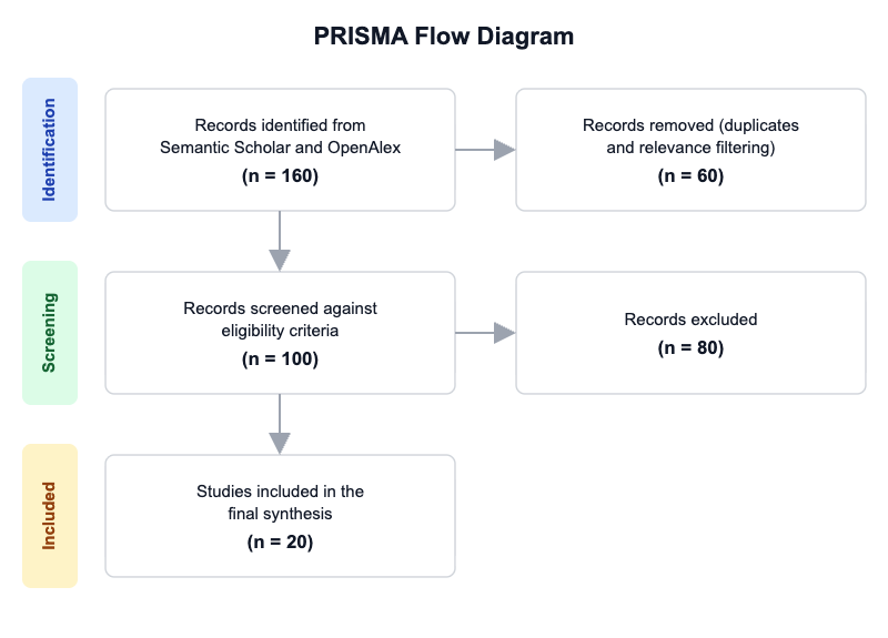
Initial database searching identified 160 records. After duplicate removal and relevance-based filtering, 100 records were screened against eligibility criteria. Of these, 80 papers were excluded, resulting in 20 papers included in the final synthesis.
PRISMA Flow Diagram
Eligibility criteria included:
- Human Studies: Does the study involve human participants or human-derived clinical data rather than only animal or cell models?
- Retinitis Pigmentosa: Does the study specifically focus on retinitis pigmentosa or a clearly labeled RP subtype/genetic form?
- Recent Work: Was the study published in 2020 or later?
- Clinical or Translational: Does the study evaluate a clinical, translational, genetic, imaging, biomarker, or therapeutic question rather than only general ophthalmology?
- Outcome Data: Does the study report at least one measurable outcome such as visual acuity, visual field, retinal structure, electrophysiology, safety, progression, or molecular endpoint?
- Novel Therapy: Does the study investigate a new or emerging therapy, gene therapy, cell therapy, device, or pharmacologic approach?
- Genetics/Biomarkers: Does the study address genotype, mutation spectrum, biomarkers, or prognostic factors?
- Review Evidence: Is the paper a review, systematic review, or meta-analysis synthesizing RP research?
All included studies met the stated eligibility criteria.
2.3 Data Extraction and Synthesis
Data extraction focused on the following variables:
- Study Focus: Extract the main research focus (e.g., gene therapy, natural history, imaging/biomarkers, pharmacologic treatment, surgical/device intervention, genetics, pathogenesis).
- Study Type: Extract the study design or publication type (e.g., clinical trial, cohort study, case series, review, systematic review, preclinical study).
- Population/Model: Extract the patient population or experimental model studied, including age group, genotype if specified, or animal/cell model.
- Intervention/Exposure: Extract the treatment, intervention, mutation, biomarker, or exposure being investigated.
- Key Outcomes: Extract the primary outcomes reported, such as visual acuity, visual fields, retinal structure, electrophysiology, safety, or progression rate.
- Main Finding: Extract the central conclusion or result of the paper in one concise sentence using only reported evidence.
- Recency/Relevance: Note why the paper is relevant to the latest research on retinitis pigmentosa, especially if it is a recent trial, novel therapy, or influential review.
Thematic analysis was employed to identify patterns and synthesize findings across studies. Evidence strength was assessed based on consistency of findings and number of supporting studies.
3. Results
3.1 Characteristics of Included Studies
| Study and Year | Study Type | Population | Intervention/Exposure | Key Focus | Outcome Measures |
|---|---|---|---|---|---|
| Cehajic-Kapetanovic et al. 2020 [1] | Phase 1/2 dose-escalation clinical trial | Patients with X-linked RP due to RPGR mutations | Subretinal AAV8-coRPGR gene therapy | First-in-human RPGR gene therapy | Safety, visual acuity, microperimetry, central retinal thickness |
| De Silva et al. 2020 [2] | Review | Clinically and genetically characterized X-linked retinal disease cohort | X-linked inheritance, gene replacement, experimental therapies | X-linked retinopathies and RPGR-related phenotypes | Clinical features, carrier phenotypes, gene-expression insights |
| Meng et al. 2020 [3] | Review | RHO-adRP context | RNA therapeutics and genome editing | Therapeutic strategies for rhodopsin-mediated RP | Potential clinical trial outcomes, visual acuity, visual fields, retinal structure |
| Botto et al. 2021 [4] | Review | Patients with inherited retinal degenerations | Gene therapy modalities | Early and late-stage gene therapy intervention | Therapeutic relevance by disease stage |
| Amato et al. 2021 [5] | Review | Broad inherited retinal disease population | Gene therapy approaches | State of the art in IRD gene therapy | Efficacy, safety, future perspectives |
| Liu et al. 2022 [6] | Review | RP as a genetic condition | Gene therapy, stem cell therapy, optogenetics, CRISPR/Cas9 | Molecular pathology and biotherapeutics | Diagnosis, treatment potential, personalized medicine |
| Cross et al. 2022 [7] | Burden review | Over a million people worldwide affected by RP | Current therapeutic landscape | Disease burden and unmet needs | Daily function, quality of life, caregiver burden |
| Cross et al. 2022 [8] | Review with qualitative physician input | RP patients across France, Germany, Italy, Spain, UK | ATMPs, gene and stem cell therapies | Current and future treatment pipeline | Development stage, accessibility, treatment stage suitability |
| Nguyen et al. 2023 [9] | Review | RP patients, variable onset and severity | Clinical management and emerging therapies | Current clinical management | Visual function, psychosocial impact, management approaches |
| Rosin et al. 2023 [10] | Review | IRD trial populations including RP | Gene therapy trial design and outcomes | Outcome measurement and trial design | Visual function, acuity, retinal structure, safety |
| Comander et al. 2023 [11] | Cohort study | Sequenced participants from vitamin A trial cohort | Vitamin A/E supplementation; genotype and ERG biomarker | Natural history and prognostic markers | Visual acuity, visual field, ERG flicker amplitude, progression rate |
| Hanany et al. 2023 [12] | Prevalence study | Multiple populations | Disease prevalence vs genetic prevalence | IRD prevalence and variant interpretation | Prevalence estimates, allele-frequency comparisons |
| Georgiou et al. 2024 [13] | Review | Diverse IRD populations | Molecular genetics, imaging, therapeutics | Phenotyping and genotyping IRDs | Clinical phenotype, imaging, therapeutic prospects |
| Karuntu et al. 2024 [14] | Review | Syndromic RP populations | Syndromic mechanisms and management | Syndromic RP spectrum | Diagnosis, clinical manifestations, management |
| Lam et al. 2024 [15] | Phase 2/3 randomized clinical trial | X-linked RP patients | Cotoretigene toliparvovec | Visual function after gene therapy | Visual acuity, retinal structure |
| Liu et al. 2024 [16] | Review | Broad RP mutation spectrum | Viral and non-viral gene therapy, gene editing | Gene therapy and genome editing | Treatment potential, mutation correction |
| Pniakowska et al. 2025 [17] | Review | RP with multiple genotypes | Luxturna, OCU400, CRISPR-Cas, MCO-010, QR-1123, SPVN06 | Genetic therapies and future directions | Toxic protein accumulation, photoreceptor preservation, cone viability |
| Ghanawi and Koch 2025 [18] | Review | RP with RPE-related and photoreceptor-related disease | AAV-mediated gene augmentation, RPE cell transplantation, pharmacologic interventions | RPE pathophysiology in RP | Morphologic alterations, therapeutic approaches |
| Reichel et al. 2025 [19] | Non-randomised controlled trial | PDE6A-associated RP | Subretinal AAV8.hPDE6A gene supplementation | Safety and vision outcomes | Visual function, central retinal thickness, visual acuity |
| Pechnikova et al. 2025 [20] | Review | Males with XLRP due to RPGR mutations | AAV5-RPGR, AGTC-501, other vector optimization | Clinical advances in XLRP gene therapy | Retinal sensitivity, visual function, gene delivery safety |
The included literature is dominated by reviews and translational papers, with a smaller number of interventional studies. The population focus is broad but converges on genetically defined RP subtypes, especially RPGR-associated X-linked disease and other mutation-specific forms. Outcome reporting is heterogeneous: some studies emphasize functional vision endpoints, whereas others center on disease burden, biomarker prediction, or molecular pathophysiology.
3.2 Thematic Findings
3.2.1 Gene therapy is the clearest translational success, but benefit is genotype- and vector-dependent
The strongest signal in the recent literature is that gene therapy has moved from conceptual promise to early clinical relevance in RP, particularly for RPGR-associated X-linked disease. In the first-in-human RPGR trial, treatment was reported as safe overall, with steroid-responsive subretinal inflammation at higher doses and visual field improvement in six patients over up to 6 months [1]. A later randomized phase 2/3 study of cotoretigene toliparvovec similarly reported potential improvement in visual function in X-linked RP, while reviews of XLRP emphasized encouraging retinal sensitivity and visual function signals from AAV-based strategies such as AAV5-RPGR and AGTC-501 [15], [20]. These findings align with the broader therapeutic logic that loss-of-function subtypes are more suitable for gene replacement than dominant gain-of-function disorders, and the X-linked literature explicitly notes this biological fit [2].
However, the evidence is not uniformly positive across genotypes. In PDE6A-associated RP, subretinal AAV8.hPDE6A did not improve visual function over 1 year and was associated with central retinal thinning and visual acuity decline [19]. That contrast suggests that therapeutic success depends not only on vector delivery but also on disease stage, target-cell viability, and gene-specific biology. Confidence: Moderate to strong for X-linked RPGR-directed approaches; limited to conflicting for broad RP gene therapy applicability.
3.2.2 The field is expanding beyond supplementation toward editing, mutation-agnostic, and rescue-based strategies
A major theme across the recent reviews is therapeutic diversification. Traditional gene supplementation remains central, but newer strategies now include RNA therapeutics, CRISPR-Cas editing, optogenetics, antisense oligonucleotides, stem cell therapy, and cone-supportive gene-independent constructs [3], [6], [17], [16]. The rationale is clear: autosomal dominant RHO-adRP involves more than 150 gain-of-function mutations, making simple gene replacement insufficient, while many patients will not qualify for mutation-specific rescue because of the breadth of genetic heterogeneity [3], [13].
The latest review literature therefore frames RP therapy as a transition from mutation replacement to multi-modal rescue of retinal function. For example, OCU400 is described as a gene-independent therapy intended to modulate retinal homeostasis across RP genotypes, MCO-010 as an optogenetic bypass of defective phototransduction, and SPVN06 as a mutation-agnostic AAV strategy delivering RdCVF/RdCVFL to support cone viability [17]. QR-1123 extends this trend by targeting a specific toxic RHO P23H variant, while CRISPR-Cas technologies remain promising but limited by off-target and delivery concerns [17]. Confidence: Moderate, because the direction of the field is consistent, but most evidence remains review-based or early-stage rather than outcome-confirmed.
3.2.3 Prognosis and trial interpretation are increasingly shaped by genotype and biomarkers rather than diagnosis alone
Recent evidence shows that RP is not a single natural history. In the genotype-stratified natural history analysis, baseline 30-Hz flicker implicit time was a strong independent predictor of progression, and genetic subtype significantly modified severity and progression patterns, extending prior findings in USH2A, RHO, RPGR, PRPF31, and EYS [11]. The same study found that vitamin E supplementation remained deleterious, while a generalized neuroprotective effect of vitamin A was not detected across all RP types [11]. This biomarker-based approach is echoed by trial-design guidance emphasizing that visual function, not acuity alone, should be assessed because acuity may miss meaningful change in field or sensitivity [10].
These data suggest that the latest RP research is moving toward patient stratification by molecular subtype and functional reserve. That shift matters because gene therapy requires surviving target cells, and retinal structure or electrophysiology may identify patients more likely to benefit than clinical diagnosis alone [11], [10]. Confidence: Strong for the prognostic value of genotype and ERG implicit time; moderate for treatment-selection implications because direct predictive validation remains limited.
3.2.4 The underappreciated role of retinal pigment epithelium and syndromic disease broadens the biological model of RP
Several recent reviews argue that RP is not solely a rod-cone photoreceptor disorder. RPE-specific mutations and secondary RPE changes are presented as important contributors to pathophysiology, with AAV-mediated gene augmentation, RPE transplantation, and pharmacologic intervention highlighted as relevant approaches [18]. This is conceptually reinforced by syndromic RP, where approximately 20–30% of cases include extra-ocular manifestations and disease mechanisms extend into ciliopathies, inherited metabolic disorders, mitochondrial disorders, and miscellaneous syndromes [14]. The implication is that a subset of patients may require systemic or multisystem evaluation rather than retina-only management.
These mechanistic and phenotypic expansions help explain why some treatments may fail even when the causal gene is known: the relevant biological target may not be limited to rods, and the disease may already involve RPE dysfunction or broader cellular networks by the time therapy is initiated [18], [14]. Confidence: Moderate, because the mechanistic narrative is consistent but mostly review-based rather than trial-confirmed.
3.2.5 Despite therapeutic progress, RP remains a major burden with limited access to curative treatment
The burden literature shows that RP continues to produce major impairment in daily functioning, employment, independence, emotional wellbeing, and caregiving capacity [7], [9]. Although voretigene neparvovec exists as a curative intervention, it applies to only 1–6% of patients according to the burden review, underscoring the narrow reach of currently approved therapy [7]. The broader therapeutic landscape therefore remains dominated by supportive care, management of complications, and trial enrollment pathways [9], [8].
This burden context is important because it shows why emerging therapies matter even when their effects are modest: functional gains in visual field, retinal sensitivity, or progression rate may translate into meaningful changes in independence for patients who otherwise have no disease-modifying options [7], [10]. Confidence: Strong for the existence of high burden and limited access to curative therapy; moderate for the size of benefit from emerging interventions.
3.3 Summary of Evidence
| Theme | Key Finding | Population Applicability | Effect Direction | Confidence Level | Supporting Studies |
|---|---|---|---|---|---|
| RPGR gene therapy shows the most convincing early clinical signal | Safety was acceptable, with steroid-responsive subretinal inflammation at higher doses and visual field improvement in six patients; later XLRP trials also reported improved retinal sensitivity/visual function | Mostly X-linked RP due to RPGR mutations; partial match to RP generally | Positive | Strong | Cehajic-Kapetanovic et al. [1], Lam et al. [15], Pechnikova et al. [20] |
| Not all gene replacement succeeds | AAV8.hPDE6A did not improve visual function over 1 year and was associated with central retinal thinning and visual acuity decline | PDE6A-associated RP; partial match to RP generally | Negative | Limited | Reichel et al. [19] |
| The therapeutic pipeline is diversifying beyond supplementation | Review-level evidence highlights RNA therapeutics, CRISPR-Cas, optogenetics, stem cells, and gene-independent rescue approaches | Broad RP populations with genotype-specific applicability | Mixed/Positive | Moderate | Meng et al. [3], Liu et al. [6], Pniakowska et al. [17] |
| Genotype and ERG biomarkers refine prognosis | Baseline 30-Hz flicker implicit time predicted progression, and vitamin E remained deleterious while vitamin A lacked generalized benefit | Sequenced RP trial cohort; direct match to RP | Mixed/Negative for vitamin E, Null for generalized vitamin A, Positive for prognostication | Strong | Comander et al. [11] |
| RP includes important syndromic and RPE-mediated biology | Approximately 20–30% of RP presents with extra-ocular manifestations, and RPE-specific mechanisms are increasingly recognized | Syndromic RP and RPE-involved RP; partial match to RP generally | Expansive/Mechanistic | Moderate | Karuntu et al. [14], Ghanawi and Koch [18] |
| The disease burden remains high and treatment access remains limited | RP severely affects daily life, employment, psychosocial wellbeing, and caregiving; approved curative therapy applies to only 1–6% | General RP population | Negative | Strong | Cross et al. [7], Nguyen et al. [9] |
4. Discussion
4.1 Principal Findings and Their Interpretation
The central message emerging from the recent RP literature is that therapeutic progress is now real but highly conditional on molecular subtype, cellular context, and disease stage. The strongest convergence is around RPGR-associated X-linked disease, where gene replacement has repeatedly shown early signals of safety and functional improvement [1], [15], [20]. This pattern is biologically plausible because RPGR-related disease is typically a loss-of-function setting in which replacement is mechanistically aligned with the causal defect [2]. By contrast, the lack of benefit in PDE6A-associated RP suggests that success is not simply a function of delivering a vector but of whether the retina still contains enough viable target tissue and whether the gene context supports rescue [19]. In other words, the field is beginning to show that “gene therapy” is not a single intervention but a family of interventions with sharply different performance depending on genetic architecture.
The biomarker literature adds an important layer of causality. Baseline ERG implicit time and genotype predicted progression, indicating that functional reserve and molecular subtype capture disease biology more accurately than diagnosis alone [11]. This strengthens the rationale for precision trial design and may explain why broad, genotype-agnostic claims of benefit remain weak. Mechanistically, recent reviews also shift the disease model beyond photoreceptors by emphasizing RPE involvement and syndromic/systemic pathways [18], [14]. That expansion matters because it helps explain why some retina-directed therapies may fail if the pathogenic process already involves RPE dysfunction or extra-retinal biology. Confidence is therefore highest for the claim that RPGR-directed therapies and genotype-based prognosis are genuinely advancing the field, while confidence is lower for generalizable claims about RP as a whole because the evidence base remains fragmented across subtypes and endpoints.
4.2 Comparison with Existing Literature and Resolution of Contradictions
The recent literature is broadly consistent with earlier expectations that RP is a heterogeneous genetic disorder, but the newer work resolves that idea into actionable therapeutic distinctions. Earlier review-level frameworks already anticipated that gene replacement would be most suitable for loss-of-function disease and that dominant gain-of-function disorders would need editing or silencing strategies [3], [6]. The latest clinical evidence supports this logic: RPGR-directed treatment appears more promising than PDE6A supplementation in the available human data [1], [19]. That consistency is meaningful because it suggests that therapeutic alignment with genetic mechanism, rather than “gene therapy” as a label, is what drives success.
The main contradiction is the coexistence of encouraging and disappointing trial outcomes. The negative PDE6A result does not negate the positive RPGR signals; rather, it likely reflects genotype-specific differences in pathobiology, stage at treatment, and possibly the adequacy of retinal target cells at the time of intervention. It may also reflect the problem that safety and efficacy can diverge: a vector can be tolerable yet still fail to rescue function if degeneration is too advanced or expression is insufficient. Another possible explanation is outcome sensitivity. Visual acuity may miss meaningful field or sensitivity changes, whereas the RPGR trial reported visual field gains and the XLRP review emphasized retinal sensitivity, suggesting that endpoint choice could partly shape perceived efficacy [10], [1], [20]. Publication bias remains a concern because reviews and pipeline papers naturally emphasize advances, but the presence of at least one clearly negative interventional study argues against an exclusively positive narrative. Methodologically, the newer emphasis on standardized visual function and biomarker endpoints appears to be a response to these contradictions and should improve comparability over time [10].
4.3 Practical Implications
For clinicians, the literature supports earlier genetic diagnosis, because therapeutic eligibility now depends increasingly on genotype, not merely phenotype [13], [11]. Patients with RPGR-associated X-linked disease currently appear to be among the most plausible candidates for emerging gene therapy trials, whereas patients with advanced structural loss or non-replaceable dominant mutations may require alternative strategies such as silencing, editing, optogenetics, or cell-based approaches [1], [17]. This argues for counseling that is specific to molecular subtype and stage of degeneration rather than generic reassurance.
At the public health level, the burden data indicate that RP remains a disabling chronic condition with limited access to curative treatment for most patients [7]. The practical implication is that health systems should not wait for a single universal therapy but should build referral pathways that enable rapid genetic testing, trial matching, and supportive low-vision care. Regulatory and reimbursement decisions should also recognize that current benefit is concentrated in narrow subgroups, so access policy must be tied to molecular eligibility and outcome measures sensitive enough to detect partial functional rescue [9], [10]. Because current evidence does not support a universal treatment threshold across RP, the appropriate response is stratified, population-specific care rather than a one-size-fits-all model.
4.4 Strengths and Limitations
A major strength of this review is that it integrates clinical trials, natural history evidence, prognostic biomarker studies, and high-level therapeutic reviews into a single synthesis of the latest RP literature. This allows interpretation across mechanistic, translational, and burden-focused domains rather than treating them separately. Another strength is that the evidence base includes both positive and negative interventional findings, which improves balance.
The included literature also has important limitations. Most evidence is either review-based or early-stage clinical data, and many studies focus on narrow genotypes such as RPGR or PDE6A rather than the broader RP population. Outcome definitions are heterogeneous, ranging from visual field and retinal sensitivity to acuity, structure, and progression biomarkers, which complicates direct comparison. Some studies address RP through proxy populations such as X-linked retinal disease or syndromic RP, which only partially matches the broader RP question. This review is also limited by reliance on the provided study data, no formal risk-of-bias assessment, and the absence of pooled quantitative meta-analysis.
5. Gaps and Future Directions
The clearest gap is the mismatch between genotype-specific success and broad RP treatment need. Future studies must move beyond RPGR-centric evidence to test whether mutation-agnostic approaches, editing strategies, or cone-supportive therapies can benefit the wider RP population [16], [17]. The negative PDE6A trial shows that efficacy cannot be assumed from preclinical success, so next-generation trials should better match disease stage, quantify remaining target tissue, and standardize outcome measures across visual function, structure, and electrophysiology [19], [10].
Another important gap is the limited integration of mechanistic biomarkers into treatment selection. The prognostic value of ERG implicit time suggests that functional biomarkers may identify patients most likely to benefit, but this has not yet been fully validated as a predictive tool for therapy response [11]. Syndromic RP and RPE-mediated disease also remain underrepresented in interventional work, despite clear evidence that extra-retinal or RPE-related biology may affect phenotype and therapeutic response [14], [18]. Finally, studies with direct comparison across delivery systems, dosing strategies, and long-term durability are needed to determine which emerging therapies can move from early promise to standard care.
6. Conclusion
The latest research on retinitis pigmentosa shows a field in transition from descriptive genetics to precision therapeutics, but the evidence supports a cautious rather than universal optimism. The most convincing advances are in genotype-defined disease, especially RPGR-associated X-linked retinitis pigmentosa, where early gene-therapy studies reported acceptable safety and functional improvement, including visual field gains in six patients [1] and encouraging retinal sensitivity/visual function signals in later XLRP trials [15], [20]. At the same time, the PDE6A trial demonstrated that not all gene replacement strategies translate into benefit, with no visual improvement over 1 year and evidence of central retinal thinning and visual acuity decline [19]. This contrast is the key lesson of the current literature: RP therapy is becoming more powerful, but also more subtype-dependent.
The broader synthesis suggests that genotype, ERG biomarkers, and retinal structural reserve now matter as much as the diagnosis itself for prognosis and trial design [11], [10]. The inclusion of syndromic disease and RPE biology further indicates that RP is mechanistically broader than a rod-cone disorder alone [14], [18]. For patients and clinicians, this means the most immediate clinical gains will likely come from earlier genetic stratification, better endpoint selection, and targeted access to emerging therapies rather than from a single universal cure. The most important unresolved question is whether mutation-agnostic or rescue-based therapies can deliver durable benefit across the genetically diverse RP population while maintaining safety.
References
- Cehajic-Kapetanovic, J., Xue, K., Martinez-Fernandez de la Camara, C., Nanda, A., Davies, A., Wood, L. J., Salvetti, A. P., Fischer, M. D., Aylward, J. W., Barnard, A. R., Jolly, J. K., Luo, E., Lujan, B. J., Ong, T., Girach, A., Black, G. C. M., Gregori, N. Z., Davis, J. L., Rosa, P. R., ... MacLaren, R. E. (2020). Initial results from a first-in-human gene therapy trial on X-linked retinitis pigmentosa caused by mutations in RPGR. Nature Medicine, 26, 354–359. https://doi.org/10.1038/s41591-020-0763-1
- De Silva, S. R., Arno, G., Robson, A. G., Fakin, A., Pontikos, N., Mohamed, M. D., Bird, A. C., Moore, A. T., Michaelides, M., Webster, A. R., & Mahroo, O. A. (2020). The X-linked retinopathies: Physiological insights, pathogenic mechanisms, phenotypic features and novel therapies. Progress in Retinal and Eye Research, 82, 100898. https://doi.org/10.1016/j.preteyeres.2020.100898
- Meng, D., Ragi, S. D., & Tsang, S. H. (2020). Therapy in rhodopsin-mediated autosomal dominant retinitis pigmentosa. Molecular Therapy, 28(10), 2139–2149. https://doi.org/10.1016/j.ymthe.2020.08.012
- Botto, C., Rucli, M., Tekinsoy, M., Pulman, J., Sahel, J.-A., & Dalkara, D. (2021). Early and late stage gene therapy interventions for inherited retinal degenerations. Progress in Retinal and Eye Research, 86, 100975. https://doi.org/10.1016/j.preteyeres.2021.100975
- Amato, A., Arrigo, A., Aragona, E., Manitto, M. P., Saladino, A., Bandello, F., & Battaglia Parodi, M. (2021). Gene therapy in inherited retinal diseases: An update on current state of the art. Frontiers in Medicine, 8, 750586. https://doi.org/10.3389/fmed.2021.750586
- Liu, W., Liu, S., Li, P., & Yao, K. (2022). Retinitis pigmentosa: Progress in molecular pathology and biotherapeutical strategies. International Journal of Molecular Sciences, 23(9), 4883. https://doi.org/10.3390/ijms23094883
- Cross, N., van Steen, C., Zegaoui, Y., Satherley, A., & Angelillo, L. (2022). Retinitis pigmentosa: Burden of disease and current unmet needs. Clinical Ophthalmology, 16, 1993–2010. https://doi.org/10.2147/OPTH.S365486
- Cross, N., van Steen, C., Zegaoui, Y., Satherley, A., & Angelillo, L. (2022). Current and future treatment of retinitis pigmentosa. Clinical Ophthalmology, 16, 2909–2921. https://doi.org/10.2147/OPTH.S370032
- Nguyen, X.-T.-A., Moekotte, L., Plomp, A. S., Bergen, A. A., van Genderen, M. M., & Boon, C. J. F. (2023). Retinitis pigmentosa: Current clinical management and emerging therapies. International Journal of Molecular Sciences, 24(8), 7481. https://doi.org/10.3390/ijms24087481
- Rosin, B., Banin, E., & Sahel, J.-A. (2023). Current status of clinical trials design and outcomes in retinal gene therapy. Cold Spring Harbor Perspectives in Medicine, 14(7), a041301. https://doi.org/10.1101/cshperspect.a041301
- Comander, J., Weigel DiFranco, C., Sanderson, K., Place, E., Maher, M., Zampaglione, E., Zhao, Y., Huckfeldt, R. M., Bujakowska, K. M., & Pierce, E. (2023). Natural history of retinitis pigmentosa based on genotype, vitamin A/E supplementation, and an electroretinogram biomarker. JCI Insight, 8(15), e167546. https://doi.org/10.1172/jci.insight.167546
- Hanany, M., Shalom, S., Ben-Yosef, T., & Sharon, D. (2023). Comparison of worldwide disease prevalence and genetic prevalence of inherited retinal diseases and variant interpretation considerations. Cold Spring Harbor Perspectives in Medicine, 14(2), a041277. https://doi.org/10.1101/cshperspect.a041277
- Georgiou, M., Robson, A. G., Fujinami, K., de Guimarães, T. A. C., Fujinami-Yokokawa, Y., Daich Varela, M., Pontikos, N., Kalitzeos, A., Mahroo, O. A., Webster, A. R., & Michaelides, M. (2024). Phenotyping and genotyping inherited retinal diseases: Molecular genetics, clinical and imaging features, and therapeutics of macular dystrophies, cone and cone-rod dystrophies, rod-cone dystrophies, Leber congenital amaurosis, and cone dysfunction syndromes. Progress in Retinal and Eye Research, 100, 101244. https://doi.org/10.1016/j.preteyeres.2024.101244
- Karuntu, J. S., Almushattat, H., Nguyen, X.-T.-A., Plomp, A. S., Wanders, R. J. A., Hoyng, C. B., van Schooneveld, M. J., Schalij-Delfos, N. E., Brands, M. M., Leroy, B. P., van Karnebeek, C. D. M., Bergen, A. A., van Genderen, M. M., & Boon, C. J. F. (2024). Syndromic retinitis pigmentosa. Progress in Retinal and Eye Research, 107, 101324. https://doi.org/10.1016/j.preteyeres.2024.101324
- Lam, B. L., Pennesi, M. E., Kay, C. N., Panda, S., Gow, J. A., Zhao, G., MacLaren, R. E., & XIRIUS Study Group. (2024). Assessment of visual function with cotoretigene toliparvovec in X-linked retinitis pigmentosa in the randomized XIRIUS phase 2/3 study. Ophthalmology, 131, 1083–1093. https://doi.org/10.1016/j.ophtha.2024.02.023
- Liu, Y., Zong, X., Cao, W., Zhang, W., Zhang, N., & Yang, N. (2024). Gene therapy for retinitis pigmentosa: Current challenges and new progress. Biomolecules, 14(8), 903. https://doi.org/10.3390/biom14080903
- Pniakowska, Z., Dzieża, N., Kustosik, N., Przybylak, A., & Jurowski, P. (2025). Genetic therapies for retinitis pigmentosa: Current breakthroughs and future directions. Journal of Clinical Medicine, 14(16), 5661. https://doi.org/10.3390/jcm14165661
- Ghanawi, H., & Koch, S. F. (2025). The versatile roles of retinal pigment epithelium in the pathophysiology of retinitis pigmentosa. Progress in Retinal and Eye Research, 108, 101390. https://doi.org/10.1016/j.preteyeres.2025.101390
- Reichel, F. F., Fischer, M. D., Stingl, K., Kuehlewein, L., Seitz, I., Peters, T., Ziegler, M., Wilhelm, B., Kohl, S., Weisschuh, N., Martus, P., Seeliger, M., Muehlfriedel, R., Paquet-Durand, F., Tsang, S. H., Bartz-Schmidt, K. U., Ueffing, M., Zrenner, E., Biel, M., Wissinger, B., & Michalakis, S. (2025). Safety and vision outcomes of subretinal gene supplementation therapy in PDE6A-associated retinitis pigmentosa: A non-randomised controlled trial. British Journal of Ophthalmology, 110(2), 173–179. https://doi.org/10.1136/bjo-2024-326480
- Pechnikova, N. A., Poimenidou, M., Iliadis, I., Zafeiriou-Chatziefraimidou, M., Iaremenko, A. V., Yaremenko, T. V., Domvri, K., & Yaremenko, A. V. (2025). Pre-clinical and clinical advances in gene therapy of X-linked retinitis pigmentosa: Hope on the horizon. Journal of Clinical Medicine, 14(3), 898. https://doi.org/10.3390/jcm14030898